A quick answer first, then details you can use. Brain MRI safety is well established in modern radiology, and Turkish hospitals follow international, standardized protocols designed to keep patients safe while capturing clear images for accurate diagnosis.
Table of Contents
Are 3T Brain MRIs Truly Safe? A Guide for Patients
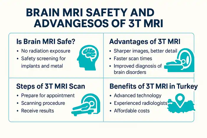
Modern MRI uses a powerful magnet and radio waves to create detailed pictures of your brain. Unlike X-rays or CT, MRI does not use ionizing radiation, which is one of the key reasons patients and clinicians consider the modality safe when appropriate screening is performed. Turkish centers that operate 3T MRI scan units apply the same global safety rules you would find in the United States or the European Union, including standardized labeling of devices as MR Safe, MR Conditional, or MR Unsafe.
The Simple Answer: Is an MRI Safe?
For the vast majority of people, yes. When facilities follow the American College of Radiology’s MR Safety guidance, MRI is considered a safe imaging test that provides critical diagnostic information without exposing you to ionizing radiation. Pre-scan screening, trained staff, and controlled access to the scanner room are core parts of that safety. If you are planning your scan in Turkey, you can expect these same safeguards. Are brain MRIs safe for everyday patients with common conditions such as headaches or dizziness? In routine practice, the benefits outweigh the minimal risks after screening.
Understanding the “No Radiation” Benefit of MRI Technology
MRI does not emit ionizing radiation. Instead, it measures signals from hydrogen atoms in your body after a radiofrequency pulse inside a strong magnetic field. This is why MRI is often selected for brain imaging, particularly in younger patients or those who require multiple follow-up scans. The lack of ionizing radiation is a primary reason clinicians consider brain MRI safety favorable compared with other modalities that do involve radiation.
What Are the Real Risks? The Power of the Magnetic Field
The risks you hear about come from the magnetic environment, not from radiation. Strong magnets can move or heat ferromagnetic objects, interfere with certain implants, and pull unsecured metal into the scanner room. Reputable hospitals manage these hazards by controlling access, screening for metal, and adhering to device-specific conditions when scanning patients with implants labeled MR Conditional. When safety rules are followed, serious events are rare.
Who Should Not Get an MRI? (Pacemakers, Implants, and Metal)
Some patients must be evaluated more carefully, especially those with cardiac pacemakers, defibrillators, neurostimulators, inner-ear implants, aneurysm clips, or metallic eye fragments. Many newer devices are MR Conditional, meaning scanning is possible when exact conditions are met such as specific field strength, head-only mode, or particular device programming. Your radiology team will verify the model and follow the conditions before proceeding. If a device is labeled MR Unsafe, MRI is generally avoided.
MRI Safety Screening in Turkish Hospitals: What to Expect
Before your appointment, you will complete a detailed safety questionnaire and speak with an MRI technologist or radiologist. Expect questions about prior surgeries, metal injuries, or implanted devices. If there is any uncertainty, staff may request operative notes, device cards, or additional checks. Facilities in Turkey that advertise 3T MRI in Turkey typically implement ACR-based workflows, which include pregnancy screening before contrast and a final metal check before entering the scan room. These standardized steps are designed to keep your experience safe from start to finish.
What is a 3T MRI? The “High-Definition” Scan Explained
A 3T MRI scan refers to a scanner with a 3-tesla magnet. In practical terms, this provides a stronger signal compared with a 1.5-tesla system and enables higher detail or faster imaging. For the brain, that can mean sharper views of small structures, subtle lesions, or microhemorrhages, which supports early and confident diagnosis.
What Does “T” in 3T Mean? (Tesla Explained)
The “T” stands for tesla, the unit that describes magnetic field strength. Higher tesla values increase the net magnetization of tissues, which improves the signal that the MRI system can detect. More signal can be traded in several ways. A radiologist may choose thinner slices, higher image resolution, or shorter scan times. This is why advantages of 3T MRI for brain imaging are often highlighted in neurology and neurosurgery settings.
3T MRI vs. Standard 1.5T MRI: A Clear Comparison
Compared with 1.5T, 3T MRI vs 1.5T MRI studies show higher signal-to-noise ratio at 3T, which can translate to crisper detail or reduced scan time. The trade-offs at 3T can include greater sensitivity to certain artifacts and potentially louder acoustic noise, all of which technologists mitigate with optimized protocols and hearing protection. In expert hands, 3T is widely considered a superior platform for many brain indications, including microlesion detection and advanced techniques.
Why a Stronger Magnet Means a Clearer Brain Scan
More signal allows the scanner to sample more information per unit time. For you, that can mean better visualization of the cortex, cranial nerves, hippocampus, and small white-matter lesions. These gains are important for conditions such as multiple sclerosis, subtle tumor margins, early ischemic changes, or pre-surgical planning where clarity matters. Patients asking about 3T MRI for brain tumor workups often benefit from this extra detail.
Is 3T MRI Always Better? When 1.5T Might Be Used
Not every clinical scenario requires 3T. Some implants are approved only under 1.5T conditions, and particular artifacts can occasionally be easier to manage at 1.5T depending on the sequence and anatomy. Radiologists in Turkey routinely decide on a case-by-case basis, balancing image quality, patient comfort, device conditions, and diagnostic needs to select the right system for you.
Why 3T MRI is the Gold Standard for Brain Imaging
The advancement from standard MRI systems to 3T MRI technology has revolutionized neurological imaging. In Turkey, modern hospitals and imaging centers have adopted 3T scanners as the gold standard for accurate, fast, and comfortable brain diagnosis. The combination of high magnetic power and improved software algorithms ensures precise detection of even the smallest structural or functional changes in the brain.
Unmatched Detail: Detecting the Smallest Brain Abnormalities
The most significant advantage of 3T MRI for brain imaging is the incredible level of detail. With twice the field strength of a 1.5T scanner, the 3T system can reveal abnormalities as small as a few millimeters. This high resolution plays a crucial role in diagnosing conditions like multiple sclerosis, vascular malformations, aneurysms, and micro-tumors that may be missed on older machines.
Radiologists can use ultra-thin slices and advanced contrast techniques, ensuring no hidden pathology is overlooked. For patients, this means earlier detection, faster intervention, and better outcomes. When it comes to brain MRI safety, having sharper images also reduces the need for repeat scans, limiting unnecessary exposure to the MRI environment.
Faster Scan Times: A Major Plus for Patient Comfort
High magnetic field strength allows for faster imaging sequences. This not only shortens the scan time but also reduces patient discomfort, especially for those who feel uneasy in confined spaces. A typical 3T MRI scan for the brain takes 15 to 25 minutes—significantly faster than older systems.
Turkish imaging centers frequently use wide-bore 3T scanners, which are more open and provide a less claustrophobic experience. Shorter scan times and comfortable design enhance patient cooperation, leading to fewer motion artifacts and clearer diagnostic images.
Advanced Neurological Diagnosis (Stroke, MS, Tumors, Epilepsy)
For neurological conditions, precision and clarity can make all the difference. The advantages of 3T MRI for brain extend beyond sharp images—they provide essential functional insights. Turkish radiology teams use 3T MRI to diagnose and monitor:
- Stroke and transient ischemic attacks (TIA): Early detection of ischemic changes helps prevent permanent damage.
- Multiple sclerosis (MS): Submillimeter lesions are more visible, improving monitoring accuracy.
- Brain tumors: 3T MRI helps define tumor boundaries and vascular supply, aiding surgeons in planning safe removal.
- Epilepsy and seizure disorders: High-resolution mapping identifies subtle cortical malformations, crucial for surgical planning.
- The combination of structural and functional MRI (fMRI) capabilities enhances the neurologist’s understanding of brain function and connectivity. These technologies support advanced procedures like brain mapping before surgery or radiation therapy.
Benefits for Functional MRI (fMRI) and Brain Mapping
Functional MRI uses changes in blood flow to map brain activity during specific tasks. The stronger signal of 3T MRI delivers higher temporal and spatial resolution, improving accuracy for detecting active brain regions. This is invaluable in preoperative planning for epilepsy, brain tumor resection, and even psychiatric research.
Turkish hospitals, especially in Istanbul and Ankara, are equipped with research-grade 3T MRI systems that combine anatomical and functional imaging. Patients benefit from this same technology used in leading European and American institutions—at a fraction of the cost. The 3T MRI in Turkey not only meets but often exceeds global imaging standards.
The 3T Brain MRI Experience in Turkey: Step-by-Step
For international patients, the process of undergoing a 3T MRI scan in Turkey is both straightforward and patient-focused. Clinics prioritize comfort, safety, and transparency, ensuring every step—from booking to receiving results—is handled with care.
How to Prepare for Your 3T MRI Appointment
Preparation for your 3T MRI for brain tumor or neurological scan is minimal. You will be asked to avoid wearing metallic objects such as jewelry, watches, or hairpins. Before the test, hospital staff will review your medical history and ensure your eligibility based on implants or other metal items.
If you are scheduled for a contrast-enhanced scan, you may be advised to fast for a few hours. Turkish radiology teams provide clear pre-scan guidance in multiple languages, ensuring smooth communication for international visitors.
Arriving at the Hospital: The Pre-Scan Process
Upon arrival, you’ll check in and fill out an MRI safety questionnaire. The staff will guide you to a changing area, where you’ll wear a comfortable gown free of metal zippers or fasteners. During the safety check, your belongings are stored outside the scanning area. The technologist will explain the procedure and answer any remaining questions about brain MRI safety.
Most Turkish hospitals emphasize patient education—technologists ensure you understand the noises, timing, and sensations you may experience, helping reduce anxiety.
Inside the 3T MRI Machine: What You Will See and Hear
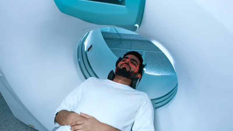
Once inside the scanner, you’ll lie on a cushioned table that slides slowly into the circular opening. You’ll hear rhythmic tapping or thumping sounds during image acquisition, which is completely normal. Earplugs or headphones are provided to reduce noise. The scan is painless, and you can communicate with the technician at any time through an intercom system.
Thanks to modern technology, today’s 3T MRI scan machines are wider, quieter, and faster than older versions.
Managing Claustrophobia: Wide-Bore Scanners and Sedation Options
If you feel anxious in confined spaces, Turkish hospitals can offer sedation or use wide-bore scanners with larger openings to create a more comfortable experience. For children or patients with severe claustrophobia, mild sedatives or open MRI options may be recommended. These practices are a crucial part of brain MRI safety, ensuring physical and emotional comfort for every patient.
After Your Scan: Getting Your Results from a Turkish Radiologist
After the scan, you can immediately resume your normal routine unless you’ve received sedation. Radiologists review and interpret your images using advanced diagnostic software. You’ll receive your results—often within 24 hours—along with a digital copy or CD for your records.
Turkish hospitals are known for combining medical precision with hospitality. The entire 3T MRI process, from appointment scheduling to results delivery, reflects world-class efficiency and patient-centered care.
Frequently Asked Questions (FAQ)
How long does a 3T brain MRI take?
Usually 15–30 minutes. The strong magnet captures clearer images faster, so your time in the scanner is shorter than older models.
Will I need a contrast dye?
Sometimes. Gadolinium contrast is used only when doctors need extra detail, such as for tumors or inflammation. It’s safe and carefully monitored.
Is the MRI noise harmful?
No. The tapping sounds come from the magnetic field changing quickly. You’ll get earplugs or headphones for comfort during the 3T MRI scan.
Can I get a 3T MRI if I’m pregnant?
Yes, in most cases. Brain MRI safety guidelines approve scans during pregnancy when necessary. Contrast dye is usually avoided unless essential.
Why choose Turkey for a 3T MRI?
3T MRI in Turkey offers top technology, expert radiologists, fast appointments, and affordable prices—all meeting international safety standards.