
Navigating the complexities of medical diagnostics can be daunting, especially when facing concerns about cancer or other serious conditions. Immunohistochemistry (IHC) is a powerful diagnostic tool that plays a crucial role in understanding diseases at the cellular level. For international patients seeking high-quality, accessible healthcare, exploring immunohistochemistry in Turkey offers a compelling combination of advanced technology, experienced specialists, and cost-effective solutions. Our institution is a leader in providing comprehensive diagnostic services, including state-of-the-art immunohistochemistry in Turkey, guiding patients towards accurate diagnoses and effective treatment pathways.
Table of Contents
What Is Immunohistochemistry (IHC) and How Does It Work?
Immunohistochemistry, often abbreviated as IHC, is a cornerstone laboratory technique used in pathology to identify specific molecules – primarily proteins or antigens – within tissue samples. Think of it as a highly specific biological ‘stain’ that reveals the ‘who’ and ‘where’ of crucial cellular components.
The core principle lies in the highly specific binding between antibodies and antigens. Antibodies, which are key players in our immune system, are designed to recognize and attach to unique targets (antigens). In IHC, scientists harness this natural ability. By applying specially prepared antibodies to a tissue sample (usually from a biopsy), pathologists can pinpoint the presence, location, and abundance of specific antigens. This information is invaluable for diagnosing diseases, especially cancer, and understanding their characteristics. Utilizing immunohistochemistry in Turkey provides access to this vital diagnostic precision.
How does it work? The “immuno” part refers to the use of antibodies. The “histo” part refers to the examination of tissues. The “chemistry” part involves the chemical reactions used to make the antibody-antigen binding visible.
In essence, an antibody designed to target a specific antigen (like a protein found only in a particular type of cancer cell) is applied to the tissue slide. If the antigen is present, the antibody binds to it. To see this binding, the antibody is linked, either directly or indirectly, to a marker – typically an enzyme. When a specific chemical (substrate) is added, the enzyme triggers a reaction, producing a colored deposit at the site of the antigen. Under a microscope, the pathologist sees this color, confirming the antigen’s presence and location. This process underpins the accuracy of immunohistochemistry in Turkey.
Immunohistochemistry vs. Immunofluorescence
While both Immunohistochemistry (IHC) and Immunofluorescence (IF) use the antibody-antigen binding principle, their key difference lies in visualization. IHC typically uses enzymes linked to antibodies, producing a colored stain visible under a standard light microscope. In contrast, Immunofluorescence uses antibodies tagged with fluorescent dyes (fluorophores). These dyes glow when exposed to specific wavelengths of light, requiring a specialized fluorescence microscope for viewing.
Here’s a quick comparison:
| Feature | Immunohistochemistry (IHC) | Immunofluorescence (IF) |
| Visualization | Colored stain (enzyme-based) | Fluorescent glow (dye-based) |
| Microscope | Standard light microscope | Fluorescence microscope |
| Signal Stability | Permanent; good for archiving | Prone to fading (photobleaching) |
| Tissue Detail | Excellent; good morphology | Can be harder to see tissue details |
| Multiplexing | Limited (usually 1-2 markers) | Higher (can view multiple markers) |
| Common Use | Routine diagnostics, cancer | Research, some diagnostics |
Key takeaway: IHC is often preferred for routine clinical diagnostics due to its permanent staining and compatibility with standard lab equipment, making it a robust choice for immunohistochemistry in Turkey. IF excels in research settings and when visualizing multiple targets simultaneously is crucial. Many top-tier labs offering immunohistochemistry in Turkey are equipped for both techniques, choosing the best method based on the diagnostic question.
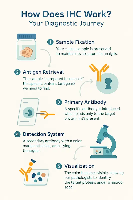
IHC Staining Procedure: Fixation, Antibody Binding & Visualization
The IHC staining procedure is a meticulous, multi-step process requiring precision and expertise to ensure accurate results. Understanding these steps helps appreciate the complexity and quality control involved in immunohistochemistry in Turkey.
- Step 1: Sample Preparation & Fixation: It begins with obtaining a tissue sample, usually through a biopsy or surgery. This tissue must be preserved quickly to prevent decay and maintain its structure. This is typically done using a chemical fixative, most commonly formalin. The fixed tissue is then embedded in paraffin wax, creating a block that can be stored and easily sliced into very thin sections (around 4-5 micrometers thick) for mounting onto microscope slides.
- Step 2: Deparaffinization & Rehydration: Before staining, the wax must be removed (deparaffinized) using solvents like xylene, and the tissue must be rehydrated through a series of alcohol solutions of decreasing concentration, ending in water.
- Step 3: Antigen Retrieval: The fixation process, while preserving structure, can sometimes mask the antigens, preventing antibodies from binding. Antigen retrieval uses heat (HIER – Heat-Induced Epitope Retrieval) or enzymes (PIER – Proteolytic-Induced Epitope Retrieval) to ‘unmask’ these antigens, making them accessible. This is a critical step for reliable IHC immunohistochemistry.
- Step 4: Blocking: Tissues can contain elements that might non-specifically bind antibodies or interfere with the detection system. Blocking steps are applied to minimize this background ‘noise’, ensuring that any signal detected is due to specific antibody-antigen binding.
- Step 5: Primary Antibody Incubation: The slide is incubated with the primary antibody – the antibody specifically designed to recognize the target antigen. This is the ‘lock and key’ moment. If the antigen is present, this antibody will bind to it.
- Step 6: Secondary Antibody & Detection System: In indirect methods (most common), a secondary antibody, which recognizes the primary antibody, is applied. This secondary antibody is usually linked to an enzyme (like HRP or AP). This step amplifies the signal. Then, a substrate/chromogen solution is added. The enzyme acts on this solution to produce a colored precipitate (e.g., brown for HRP/DAB) exactly where the primary antibody has bound.
- Step 7: Counterstaining & Mounting: To provide context and visualize the tissue structure, a counterstain (like hematoxylin, which stains nuclei blue) is often applied. Finally, the slide is dehydrated, cleared, and a coverslip is mounted, making it a permanent record ready for microscopic examination. The quality of immunohistochemistry in Turkey relies heavily on the skilled execution of each step.
When and Why Is IHC Performed?
Immunohistochemistry is not a routine test for every patient; it’s a specialized tool used when specific questions need answering, particularly after initial tests like a standard biopsy review. Its ability to identify specific proteins makes it indispensable in various medical scenarios.
Quick list: IHC is primarily performed to:
- Diagnose and Classify Cancers: This is the most common use. IHC can differentiate between types of cancer that look similar under a standard microscope (e.g., distinguishing between different types of lung cancer or lymphoma). It can also help determine if a tumor is benign or malignant.
- Identify the Origin of Metastatic Cancer: When cancer spreads (metastasizes), it’s often crucial to find its original source (the primary tumor) to guide treatment. IHC can identify markers characteristic of specific organs, helping to pinpoint where the cancer began. This is a vital application of immunohistochemistry in Turkey for complex cancer cases.
- Predict Prognosis and Treatment Response: IHC can detect proteins that indicate how aggressive a cancer might be (prognostic markers) or predict whether a cancer will respond to specific treatments (predictive markers). For example, testing for estrogen receptors (ER) and progesterone receptors (PR) in breast cancer helps determine if hormone therapy will be effective.
- Detect Infectious Agents: IHC can be used to identify specific viruses, bacteria, or parasites within tissues when other methods are inconclusive.
- Diagnose Certain Neurological and Muscular Disorders: As we’ll explore later, IHC helps identify abnormal protein deposits or deficiencies in conditions like Alzheimer’s disease or muscular dystrophy.
Key takeaway: IHC is performed when detailed information about the proteins within cells is needed to make a precise diagnosis, determine a patient’s outlook, or select the most effective treatment. Its role in personalized medicine is growing, making access to high-quality immunohistochemistry in Turkey increasingly important for patients worldwide.
Diagnostic Applications of IHC: From Cancer to Neurological Disorders
Immunohistochemistry (IHC) serves as a critical diagnostic bridge, translating the visual patterns under a microscope into specific molecular diagnoses. Its applications span a wide spectrum of diseases, offering insights that are often unattainable through other methods. The primary power of IHC lies in its ability to pinpoint specific proteins within the context of tissue architecture, providing crucial clues for diagnosis, prognosis, and treatment selection. For international patients, accessing advanced diagnostic immunohistochemistry in Turkey means tapping into a wealth of expertise across various medical fields, from oncology to neurology, ensuring a comprehensive understanding of their health status.
The utility of IHC immunohistochemistry is vast. It’s not just about confirming a diagnosis; it’s about refining it. Is a tumor cancerous or benign? If cancerous, what specific type is it, and where did it originate? What are its biological drivers, and which therapies are most likely to work? These are the questions that IHC helps answer. Furthermore, its role extends beyond cancer, aiding in the identification of infectious agents and the characterization of degenerative diseases affecting the brain and muscles. Pursuing immunohistochemistry in Turkey provides patients with a reliable pathway to obtaining these vital answers.
Identifying Cancer Subtypes & Differentiating Benign vs. Malignant Tumors
Cancer diagnosis is where immunohistochemistry truly shines and is most frequently applied. While a traditional biopsy (H&E stain) can reveal abnormal cell growth, IHC provides a deeper, molecular layer of information. It is indispensable for accurately classifying tumors, which is fundamental for planning treatment. For instance, different types of lung cancer (like adenocarcinoma versus squamous cell carcinoma) might look similar but express different proteins. IHC tests can identify these specific protein markers, leading to a precise classification and, consequently, more targeted and effective therapies. The expertise in immunohistochemistry in Turkey is particularly strong in the field of oncology.
One of the most critical roles of IHC is differentiating between benign (non-cancerous) and malignant (cancerous) tumors, especially when routine staining is ambiguous. Certain IHC markers are strongly associated with malignancy, while others indicate benign growth. Furthermore, when cancer has spread (metastasized), IHC can act like a ‘cellular GPS’. By testing the metastatic tumor for proteins characteristic of different organs (e.g., PSA for prostate, TTF-1 for lung, GATA3 for breast), pathologists can often determine the primary site of the cancer, even if it wasn’t initially known. This is a crucial application of immunohistochemistry in Turkey for complex cancer cases.
Quick list: Key IHC applications in cancer:
- Tumor Classification: Identifying specific subtypes (e.g., breast cancer: ER/PR/HER2 status; lymphomas: B-cell vs. T-cell).
- Benign vs. Malignant: Differentiating borderline or uncertain growths.
- Metastasis Origin: Identifying the primary source of secondary tumors.
- Prognostic Markers: Assessing tumor aggressiveness (e.g., Ki-67 proliferation index).
- Predictive Markers: Determining likely response to targeted therapies (e.g., PD-L1 for immunotherapy).
Detecting Neurological Diseases: Alzheimer’s, Parkinson’s & Muscular Dystrophy
While most associated with cancer, IHC is also a valuable tool in diagnosing certain neurological and neuromuscular disorders. These conditions often involve the abnormal accumulation or absence of specific proteins in the brain or muscle tissue. IHC allows pathologists to visualize these protein changes directly, often confirming a diagnosis that might be suspected based on clinical symptoms but requires tissue evidence. Leading centers for immunohistochemistry in Turkey collaborate closely with neurologists to provide these specialized tests.
- Alzheimer’s Disease: IHC can detect the hallmark protein deposits in the brain associated with Alzheimer’s – amyloid-beta plaques and tau tangles. While brain biopsies for this purpose are rare in living patients, IHC is crucial for confirming the diagnosis during post-mortem examinations, which helps in research and understanding the disease.
- Parkinson’s Disease: Similarly, IHC can identify Lewy bodies, which are abnormal aggregates of the protein alpha-synuclein found in specific brain regions of individuals with Parkinson’s disease and other synucleinopathies. Again, this is primarily used in post-mortem analysis for definitive diagnosis.
- Muscular Dystrophy: In various forms of muscular dystrophy, specific proteins essential for muscle structure and function are either absent or deficient. IHC performed on a muscle biopsy can identify these protein deficiencies (like dystrophin in Duchenne muscular dystrophy), providing a definitive diagnosis and helping to classify the specific type of dystrophy. Accessing reliable muscle biopsy analysis, including immunohistochemistry in Turkey, is vital for these patients.
Research & Hematology-Oncology Uses of IHC
Beyond routine clinical diagnostics, immunohistochemistry is an indispensable tool in medical research. Scientists use IHC to understand disease mechanisms, identify new diagnostic markers, test the effects of potential drugs, and explore cellular pathways. Many breakthroughs in our understanding of cancer, neurodegeneration, and infectious diseases have relied heavily on IHC experiments. The dynamic medical research community in Turkey frequently employs IHC, contributing to global knowledge. The availability of advanced immunohistochemistry in Turkey supports both clinical practice and research endeavors.
In hematology-oncology – the field dealing with cancers of the blood, bone marrow, and lymph nodes – IHC is critically important. Diagnosing lymphomas and leukemias often requires identifying specific cell surface markers (CD markers) to determine the exact lineage and subtype of the cancerous cells.
While flow cytometry is often used for liquid samples, IHC is essential for examining lymph nodes and bone marrow biopsies. It helps differentiate reactive (inflammatory) processes from cancerous ones and classifies lymphomas with high precision, guiding complex treatment decisions. For patients seeking specialized care, the capabilities of immunohistochemistry in Turkey in hematology-oncology are a significant advantage.
Turkey’s Strengths in Immunohistochemistry
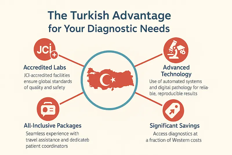
Turkey has rapidly emerged as a leading destination for medical tourism, and its capabilities in advanced diagnostics like immunohistochemistry (IHC) are a significant draw. Patients seeking immunohistochemistry in Turkey find a healthcare system that combines international standards of quality with exceptional value. The nation’s investment in modern medical infrastructure, coupled with a deep pool of skilled professionals, creates an environment where complex diagnostic procedures like IHC are performed with precision and care. Choosing Turkey means accessing a healthcare ecosystem dedicated to excellence, making it a wise choice for your diagnostic needs.
Key takeaway: Turkey offers a compelling package for patients requiring IHC: internationally accredited laboratories, highly experienced pathologists, advanced technology, and a commitment to personalized patient care. This ensures that when you opt for immunohistochemistry in Turkey, you are receiving world-class diagnostic services. Our institution stands at the forefront of this, providing seamless care for international visitors.
Accredited Pathology Labs in Istanbul and Nationwide
A cornerstone of reliable diagnostics is the quality of the laboratory where tests are processed. Turkey boasts a significant number of pathology laboratories, particularly in major cities like Istanbul, Ankara, and Izmir, that meet and often exceed global standards. Many leading hospitals and private laboratories hold prestigious international accreditations, such as those from the Joint Commission International (JCI).
This accreditation signifies a commitment to the highest levels of patient safety and quality of care, covering everything from sample handling to reporting procedures. When considering immunohistochemistry in Turkey, choosing a JCI-accredited facility provides an extra layer of confidence.
These accredited labs are equipped with cutting-edge technology. Automated IHC stainers, digital pathology platforms for easier consultation and archiving, and a comprehensive menu of available antibody tests ensure that pathologists have the best tools at their disposal. Strict internal and external quality control programs are standard practice, guaranteeing the accuracy and reproducibility of results. This technological prowess means that immunohistochemistry in Turkey is performed using the same, and sometimes even more advanced, methods found in leading Western healthcare systems. Our facility prides itself on maintaining a state-of-the-art laboratory, ensuring top-tier diagnostic services.
Expertise of Turkish Pathologists & Multidisciplinary Teams
Technology alone isn’t enough; the human element – the expertise of the pathologist – is paramount in IHC. Turkish pathologists are highly trained professionals, many of whom have received education or specialized training in Europe and the United States. They possess deep experience in interpreting complex IHC slides across various specialties, particularly in oncology and hematology. Their proficiency ensures that the subtle nuances of IHC staining are accurately interpreted, leading to a precise diagnosis. When seeking the best doctor for immunohistochemistry in Turkey, patients will find a wealth of experienced specialists.
Furthermore, healthcare in leading Turkish institutions operates on a multidisciplinary team (MDT) model. This means that pathologists work in close collaboration with oncologists, surgeons, radiologists, and other specialists. IHC results are not viewed in isolation; they are discussed within the MDT to formulate a comprehensive understanding of the patient’s condition and to develop the most appropriate treatment plan. This collaborative approach ensures that the findings from immunohistochemistry in Turkey are translated into actionable and effective clinical decisions, providing patients with integrated and holistic care. Our MDTs ensure every aspect of your diagnosis is considered.
Gender-Specific and Personalized IHC Approaches
The era of one-size-fits-all medicine is fading, replaced by a focus on personalized and precision medicine. Immunohistochemistry is a key enabler of this shift, and Turkey’s healthcare system is embracing this approach. By identifying specific molecular markers, IHC allows for treatments to be tailored to the individual characteristics of a patient’s disease. This is particularly evident in cancer treatment, where IHC results guide the use of targeted therapies and immunotherapies, improving outcomes and minimizing side effects. Opting for immunohistochemistry in Turkey means benefiting from this personalized approach.
Turkish medical centers are also increasingly aware of the importance of gender-specific medicine. While the IHC techniques themselves are standard, the interpretation and application of results can sometimes be viewed through a lens that considers potential differences between genders in disease presentation and response. For instance, the IHC panels for breast cancer (ER, PR, HER2) are fundamentally gender-specific, but awareness extends to other areas as well. This commitment to a nuanced, patient-centric approach means that immunohistochemistry in Turkey aims to provide the most relevant and effective diagnostic information for every individual, ensuring that care is not just advanced, but also deeply personal.
Immunohistochemistry Costs & Packages in Turkey
One of the most compelling reasons international patients choose Turkey for medical procedures is the significant cost advantage, especially when compared to North America and Western Europe. This holds true for advanced diagnostics like immunohistochemistry (IHC). Pursuing immunohistochemistry in Turkey allows patients to access world-class diagnostic technology and expertise at a fraction of the price they might pay in their home countries. Importantly, this affordability does not come at the expense of quality. Strict accreditation standards, modern facilities, and experienced specialists ensure that the value offered by immunohistochemistry in Turkey is matched by its reliability and precision.
Key takeaway: Turkey presents a highly attractive option for patients needing IHC, offering substantial cost savings compared to the West without compromising on the quality of diagnostics, technology, or medical expertise. Our institution provides transparent pricing and comprehensive packages, making the process of obtaining immunohistochemistry in Turkey both affordable and straightforward for international visitors. We are committed to delivering exceptional value alongside exceptional care.
Cost Comparison: Turkey vs. Europe/USA
The difference in cost for medical services between Turkey and countries like the USA, Canada, UK, or Germany can be striking. For immunohistochemistry, patients can often expect savings ranging from 50% to 70%, and sometimes even more. While an IHC test panel might cost thousands of dollars or euros in the West, the immunohistochemistry costs in Turkey are significantly lower. For example, a standard IHC workup for a cancer biopsy might range from $300 to $800 USD (or equivalent in EUR) in Turkey, whereas the same panel could easily exceed $1,500 to $4,000 USD in the United States or Western Europe.
This price difference is due to several factors, including lower operational costs, favorable exchange rates, and a government focus on promoting medical tourism. However, it’s crucial to understand that these savings are achieved while maintaining high standards. Turkish labs use the same high-quality reagents, antibodies, and automated equipment as their Western counterparts. The significant cost advantage makes high-level diagnostics, including comprehensive immunohistochemistry in Turkey, accessible to a much broader range of global patients who might otherwise face financial barriers to obtaining an accurate diagnosis.
Factors Influencing IHC Pricing
While immunohistochemistry in Turkey is generally affordable, the exact cost can vary based on several key factors. Understanding these can help patients anticipate their expenses:
- Number and Type of Antibodies: This is the most significant factor. Each IHC test uses a specific antibody to detect a particular protein. Some diagnostic workups require only one or two antibodies, while others, like classifying complex lymphomas or undifferentiated tumors, might need a panel of ten, fifteen, or even more. The more antibodies used, the higher the cost. Some specialized antibodies are also more expensive than common ones.
- Complexity of the Case: Cases requiring extensive investigation, multiple tissue blocks, or second opinions from multiple pathologists may incur higher fees.
- Laboratory Fees: This covers the cost of materials (slides, reagents, fixatives), equipment use, and the technical staff’s time in performing the IHC staining and processing.
- Pathologist’s Fees: This covers the professional time and expertise of the pathologist who examines the slides, interprets the results, and writes the comprehensive diagnostic report. Experienced, highly specialized pathologists may command higher fees.
- Hospital or Clinic Choice: Prices can vary slightly between different hospitals and private labs, though competition generally keeps immunohistochemistry costs competitive across accredited institutions.
- Urgency: Expedited or ‘STAT’ processing, if available and requested, may carry an additional charge.
Despite these variables, the overall cost structure for immunohistochemistry in Turkey remains significantly more favorable than in many other developed nations. Our patient coordinators provide clear, upfront cost estimates.
All-Inclusive Diagnostic Packages for International Patients
To simplify the process for overseas patients, many leading Turkish healthcare providers, including our institution, offer all-inclusive diagnostic packages. These packages are designed to provide a seamless and stress-free experience, bundling various costs and services into a single, transparent price. This approach helps patients budget effectively and avoids unexpected expenses when seeking immunohistochemistry in Turkey.
A typical diagnostic package might include:
- The IHC tests themselves (often defined up to a certain number of antibodies, with clear pricing for additional markers if needed).
- The pathologist’s consultation and reporting fees.
- Consultations with the referring specialist (e.g., oncologist or surgeon) before and after the test.
- Translation services if required.
- Assistance with travel logistics, such as airport transfers and hotel recommendations or bookings.
- Dedicated patient coordinators to guide you through every step.
These packages transform the process of obtaining immunohistochemistry in Turkey from a purely medical procedure into a well-managed healthcare journey. They emphasize patient comfort and convenience, allowing you to focus on your health while we handle the details. We encourage you to inquire about our tailored diagnostic packages designed specifically for international patients.
Quality Assurance & Technological Advances in IHC
Achieving an accurate diagnosis through immunohistochemistry (IHC) hinges on meticulous attention to detail and rigorous quality control at every stage. Inaccurate or unreliable results can lead to misdiagnosis and inappropriate treatment, making quality assurance (QA) paramount. Leading healthcare institutions offering immunohistochemistry in Turkey understand this critical need and have implemented robust QA programs and embraced technological advancements to ensure the highest standards of diagnostic precision. This commitment guarantees that patients receive dependable results they can trust.
Key takeaway: The reliability of immunohistochemistry in Turkey is built upon a strong foundation of international-standard quality assurance protocols, from initial sample handling to final interpretation, complemented by the adoption of cutting-edge technologies. Our facility prioritizes these measures to deliver consistently accurate and reproducible diagnostic information, underpinning our commitment to patient safety and effective care. When you choose us for immunohistochemistry in Turkey, you choose a service dedicated to quality.
Sample Preservation and Error-Mitigation Strategies
The journey of an IHC test begins long before the slide reaches the microscope; it starts with the tissue biopsy. The ‘pre-analytical’ phase – how the tissue is obtained, handled, and preserved – is arguably the most critical and can significantly impact the final result. Improper fixation (using the wrong fixative, incorrect duration, or delayed fixation) can damage tissue structure or mask antigens, making accurate IHC immunohistochemistry impossible. Reputable centers for immunohistochemistry in Turkey adhere to strict, standardized protocols for sample handling.
Quick list: Key error-mitigation strategies include:
- Standardized Fixation: Using 10% neutral buffered formalin for specific durations, based on tissue size and type.
- Proper Tissue Processing: Careful dehydration, clearing, and paraffin embedding to maintain tissue integrity.
- Unique Patient Identifiers: Rigorous labeling and tracking systems using barcodes or similar methods to prevent sample mix-ups.
- Optimized Sectioning: Cutting thin, consistent tissue sections to ensure even staining and optimal visualization.
- Controlled Storage: Maintaining appropriate temperature and humidity for stored tissue blocks and slides.
By controlling these pre-analytical variables, top Turkish labs minimize the risk of errors that could compromise the diagnostic value of immunohistochemistry in Turkey. Our laboratory follows stringent international guidelines for every sample we process.
Ensuring Reliable, Reproducible Results
Once a sample is properly prepared, the staining process itself requires strict standardization and control to ensure results are both reliable (accurate) and reproducible (consistent across different runs and different labs). This involves several key QA measures integrated into the workflow for immunohistochemistry in Turkey.
- Validated Antibodies & Protocols: Only antibodies that have been thoroughly tested and validated for their specific targets and applications are used. Staining protocols are optimized and standardized for each antibody.
- Use of Controls: Every IHC run includes controls. Positive controls (tissue known to contain the target antigen) confirm that the staining process worked correctly. Negative controls (tissue known not to contain the antigen, or slides processed without the primary antibody) ensure that any staining observed is specific and not due to background noise or non-specific binding.
- Automated Staining Platforms: Many leading labs offering immunohistochemistry in Turkey utilize automated IHC stainers. These machines provide highly consistent and reproducible staining by controlling variables like incubation times, temperatures, and reagent dispensing, reducing the potential for human error inherent in manual methods.
- Proficiency Testing: Reputable labs participate in external quality assurance (EQA) or proficiency testing programs. They receive ‘unknown’ samples from an external body and are graded on their ability to achieve the correct results, providing an objective measure of their performance.
- Standardized Interpretation: Pathologists follow established guidelines and scoring systems when interpreting IHC results, promoting consistency in how stains are read and reported.
These measures collectively ensure that the immunohistochemistry in Turkey you receive meets high standards of quality, providing a trustworthy basis for clinical decisions.
Emerging Technologies and Future Directions in IHC
Immunohistochemistry is not a static field; it is constantly evolving. Turkish diagnostic centers are keen adopters of emerging technologies that enhance the power and precision of IHC. These advancements are shaping the future of pathology and improving patient care.
- Digital Pathology: This involves scanning stained slides at high resolution to create digital images. These images can be viewed, managed, and analyzed on a computer. Digital pathology facilitates easier consultations (telepathology), allows for powerful image analysis algorithms, and creates a permanent, non-degrading digital archive. Many centers for immunohistochemistry in Turkey are integrating digital pathology.
- Quantitative Image Analysis: Software can now analyze digital IHC slides to provide objective, quantitative data – for example, precisely measuring the percentage of cells positive for a marker or the intensity of the stain. This moves beyond subjective interpretation, increasing accuracy and reproducibility.
- Multiplex IHC (mIHC) / Immunofluorescence (mIF): These advanced techniques allow pathologists to visualize multiple markers (sometimes 5, 10, or even more) on a single tissue section. This provides a much deeper understanding of the cellular landscape, cell-to-cell interactions, and the tumor microenvironment, which is particularly important for immunotherapy research and application. Leading research and diagnostic labs in Turkey are exploring these powerful tools.
These innovations demonstrate that choosing immunohistochemistry in Turkey means accessing not only established best practices but also the future directions of diagnostic pathology, ensuring patients benefit from the very latest advancements in the field.
Patient Guide & Frequently Asked Questions
Embarking on a medical journey abroad, even for diagnostics, involves many questions. This guide aims to answer common queries for international patients considering immunohistochemistry in Turkey, providing practical information to help you plan and feel confident in your choice. Understanding the process, timelines, and how to select the right provider is key to a smooth and successful experience. Our institution is dedicated to supporting international patients, ensuring clarity and comfort throughout their diagnostic journey for immunohistochemistry in Turkey.
Key takeaway: Planning for immunohistochemistry in Turkey is straightforward. Understanding the test timeline, how results are interpreted, and how to select the best facility will empower you. We offer comprehensive support and clear communication to guide you every step of the way, ensuring your experience is as seamless as possible.
How Long Does an IHC Test Take?
A common question patients ask is: “How long until I get my results?” The turnaround time (TAT) for an IHC test can vary, but generally, you can expect results within a few days to about a week after the tissue sample (biopsy) arrives at the pathology lab. It’s important to understand that immunohistochemistry is a multi-step process, and each step takes time to perform correctly.
Here’s a typical timeline:
- Biopsy & Transport (Variable): This depends on when your biopsy is taken and how long it takes to reach the pathology lab. For international patients, coordinating this might involve sending existing biopsy blocks or travelling to Turkey for the procedure.
- Fixation & Processing (12-24 hours): Once in the lab, if it’s fresh tissue, it needs to be fixed and then processed into a wax block. This is crucial for preservation.
- Sectioning & Mounting (Few hours): The wax block is sliced very thinly, and the sections are mounted onto glass slides.
- IHC Staining (4-8 hours to 1 day): This involves deparaffinization, antigen retrieval, antibody incubation, and visualization. While automated systems speed this up, it still requires several hours. The complexity and number of antibodies for your immunohistochemistry in Turkey test will influence this step.
- Interpretation & Reporting (1-3 days): The pathologist examines the stained slides under a microscope, interprets the findings in the context of your clinical history, and writes a detailed report. Complex cases may require consultation or additional stains, extending this time.
- Report Delivery (Few hours): Once finalized, the report is sent to your referring physician and made available to you.
Key takeaway: While the core IHC staining can be done relatively quickly, the entire process from sample receipt to final report typically takes 3 to 7 working days. At our facility providing immunohistochemistry in Turkey, we strive for efficiency while never compromising the quality and accuracy required for these crucial tests.
How to Read and Interpret IHC Results
Receiving a pathology report filled with medical terms can be intimidating. While your doctor is the best person to explain the results in detail, understanding the basics can be helpful. An IHC report essentially describes what proteins were found, where they were found in the cells, and how much was present.
Here’s how to understand common terms:
- Positive (+): This means the antibody ‘stuck’ to its target antigen, indicating the protein is present in your cells.
- Negative (-): This means the antibody did not find its target protein.
- Staining Pattern: Describes where the stain is seen (e.g., nucleus, cytoplasm, cell membrane). This is important because a protein’s location can affect its function.
- Staining Intensity: Describes how strong the color is (e.g., weak/1+, moderate/2+, strong/3+). This gives an idea of the amount of protein.
- Percentage of Positive Cells: Indicates what proportion of the relevant cells (e.g., tumor cells) are showing the stain.
How to interpret immunohistochemistry results? It’s crucial to remember that IHC results are not a diagnosis in isolation. The pathologist combines these findings with the standard H&E slide review and your clinical history. For example, a “positive” result for ER in breast cancer is good news for hormone therapy, while a “positive” result for a specific marker in an unknown tumor helps identify its origin. When you undergo immunohistochemistry in Turkey, our specialists ensure that reports are comprehensive and are happy to facilitate clear discussions about their meaning with your primary doctor.
Choosing the Best Doctor or Clinic for IHC in Turkey
Selecting the right facility is vital. When looking for the best clinic for immunohistochemistry in Turkey or the best doctor for immunohistochemistry in Turkey, consider these factors:
- Accreditation: Look for internationally recognized accreditations like JCI. This is a strong indicator of quality and safety standards.
- Laboratory Technology: Does the facility use modern, automated IHC equipment and offer a wide range of antibodies?
- Pathologist Expertise: Inquire about the experience and sub-specialty training of the pathologists, especially in areas relevant to your condition (e.g., oncology, neurology).
- Multidisciplinary Approach: Does the clinic or hospital facilitate collaboration between pathologists and clinicians?
- International Patient Services: Do they have experience handling international patients? Look for services like English-speaking staff, translation, package deals, and travel assistance. This is particularly important for patients travelling from nearby regions like Azerbaijan or further afield.
- Turnaround Time & Communication: Ask about their typical TAT and how they communicate results. Clear, prompt communication is essential.
- Cost Transparency: Ensure they provide clear, upfront pricing and detailed explanations of what is included.
- Patient Reviews & Testimonials: Look for feedback from other international patients.
Key takeaway: Our institution excels across these criteria, offering JCI-accredited services, advanced technology, highly experienced pathologists, and dedicated support for international patients. We are a leading choice for those seeking reliable, high-quality, and cost-effective immunohistochemistry in Turkey. Contact our international patient department today to learn how we can assist you.
A precise diagnosis is the crucial first step toward recovery, and an immunohistochemistry in Turkey report provides the detailed map needed to plan your therapeutic journey. Once your diagnostic results are clear, the next step is to explore the most effective treatment options tailored to your specific condition.
Our institution offers a comprehensive range of advanced therapeutic services that align with your diagnostic findings. We encourage you to learn more about our world-class treatment programs, including state-of-the-art options such as Immunotherapy with Keytruda (Pembrolizumab) for various cancers, specialized Lymphoma Chemotherapy regimens, intricate Brain AVM Treatment, and life-saving Pediatric Bone Marrow Transplantation. Each of these pathways is supported by our multidisciplinary teams dedicated to achieving the best possible outcomes for our patients.
