

A brain MRI is one of the most advanced diagnostic tools in modern medicine, offering a precise, noninvasive look inside your head. Knowing how to prepare for brain MRI can make the process comfortable and stress-free. This guide explains every step — from understanding what a brain scan shows to knowing exactly what to expect on the day of your procedure.
Table of Contents
What Is a Brain MRI and Why Might You Need One?
Magnetic resonance imaging, commonly called brain MRI, uses a powerful magnetic field and radio waves to produce detailed images of your brain and surrounding structures. Unlike CT scans, MRI does not use radiation. It is often performed when doctors need clearer images of soft tissues to evaluate neurological symptoms or monitor existing conditions.
A Simple Guide: How MRI Technology Sees Your Brain (No Radiation)
MRI technology works by aligning hydrogen atoms in your body with a magnetic field. When radiofrequency pulses are applied, these atoms produce signals that are transformed into images by the computer. This allows radiologists to see even the smallest abnormalities in the brain’s tissue, blood vessels, or nerves. Whether you are getting a head MRI or a specialized functional MRI, the process is completely safe and painless.
Common Reasons Your Doctor Ordered a Head Scan
There are several reasons why your doctor may recommend a brain MRI. The scan can help detect causes of chronic headaches, dizziness, seizures, or vision problems. It is also commonly used to assess stroke damage, brain tumors, infections, and multiple sclerosis. In some cases, it helps evaluate memory loss, trauma, or unexplained neurological changes. Understanding these reasons for brain MRI can reduce anxiety by showing that the test is a standard, informative part of medical care.
Before moving on to how to prepare for brain MRI, it is helpful to understand that every scan is customized to the patient. Some people need a basic head MRI, while others undergo an advanced functional MRI (fMRI) to measure brain activity. Your doctor will choose the right type depending on your symptoms and medical history.
How to Prepare: Your Complete Brain MRI Checklist
Proper preparation can make your scan more effective and comfortable. The following MRI preparation checklist covers everything from what to tell your doctor to what you should wear.

Before Your Appointment: What to Tell Your Doctor
To prepare for brain MRI safely, inform your doctor about any medical implants, metal fragments, pacemakers, or tattoos that contain metallic ink. Discuss any allergies, especially if a contrast dye may be used. Patients with metal implants or certain medical devices may need additional screening or an alternative imaging method. It is also important to mention if you are pregnant or breastfeeding, as this might influence how the scan is conducted.
The Day of Your Scan: What to Eat, Drink, and Wear
Generally, you can eat and drink normally before your MRI unless told otherwise. For some procedures involving contrast dye, fasting for a few hours may be required. Wear loose, comfortable clothing without metal zippers or buttons. If your facility requires it, you will change into a hospital gown before entering the scan room. Avoid wearing jewelry, makeup with metallic particles, or hair accessories, as they can interfere with the magnetic field.
The “No Metal” Rule: What You Must Leave at Home
MRI machines are highly sensitive to metal, so following the “no metal” rule is essential. Before entering the scan area, remove all jewelry, watches, hearing aids, credit cards, and phones. Even small metallic items can distort the image or pose a safety risk. Patients with metal implants should double-check with the radiologist, as most modern implants are MRI-compatible, but it is vital to confirm in advance.
The Brain MRI Procedure: A Step-by-Step Walkthrough
Once you have completed the MRI preparation checklist, it is time to understand what actually happens during the scan. Knowing each step of the MRI procedure helps you stay calm and confident. From check-in to image completion, the entire process is designed to be safe, comfortable, and precise.
Step 1: Checking In and Changing into a Gown
When you arrive at the imaging center, a receptionist or radiology nurse will review your medical information and confirm your safety questionnaire. You may be asked again about any metal implants, piercings, or devices. After this, you will be directed to a changing area. Most facilities provide a gown to eliminate any risk of metallic clothing parts interfering with the scan. Lockers are available for storing your belongings.
If you have brought previous imaging results or doctor’s notes, hand them to the technologist before starting. They help compare current images with earlier results for a more accurate evaluation.
Step 2: Meeting the Technologist and Final Safety Screen
Next, the MRI technologist introduces themselves and explains the procedure. This is a good time to ask questions about how to prepare for brain MRI or clarify any concerns about the scanner environment. A final safety check will ensure that you have removed every metal item, including hearing aids, watches, and credit cards. The technologist will also confirm if you need contrast dye and whether an IV line should be placed before scanning.
Throughout the process, patient safety is the top priority. The MRI staff are trained to recognize contraindications and adapt settings for people with pacemakers, joint replacements, or other implants.
Step 3: Getting Positioned on the Scanner Table
After the safety screening, you will lie down on a sliding table that moves into the scanner tunnel. A soft coil, shaped like a helmet or head frame, will be placed around your head. This device helps capture high-resolution images. The technologist adjusts padding and cushions to keep you comfortable and ensure your head remains completely still.
Some facilities offer earplugs or headphones to reduce the sound of the machine. You may also be given a small squeeze ball or call button that lets you communicate instantly if you feel discomfort.
Step 4: Inside the Scanner: What It Feels and Sounds Like
As the scan begins, the table moves smoothly into the magnet. The scanner makes rhythmic tapping and humming noises as it collects data from different angles. These sounds are normal and indicate that the magnetic field is changing rapidly to capture detailed images of your brain structures.
Although the machine may appear tight, the process is entirely painless. The key to accurate imaging is staying still throughout the scan. If you feel anxious, you can close your eyes and focus on breathing slowly and evenly. Many patients find that visualizing calm or listening to music through MRI-safe headphones helps them relax.
Step 5: Staying Still and Communicating (The Call Button)
During the brain MRI, the technologist will monitor you from an adjacent control room and communicate through an intercom. They will provide gentle reminders to stay still between scans and inform you how long each sequence will take. If you experience any sudden discomfort or anxiety, pressing the call button immediately pauses the procedure so the technologist can assist you.
The entire scan typically lasts between 30 and 60 minutes, depending on whether contrast dye is used. The key is patience and cooperation — remaining still ensures sharper, more diagnostic images.
Understanding MRI with Contrast (Gadolinium)
Some patients require a contrast-enhanced MRI to highlight specific brain tissues or detect small abnormalities that a standard scan might miss. This involves injecting a small amount of gadolinium-based dye into a vein.
What Is Contrast Dye and Why Is It Used?
Contrast dye helps radiologists see clearer distinctions between normal and abnormal brain tissue. It makes tumors, blood vessel malformations, or areas of inflammation appear more visible on the scan. Not every patient needs contrast; your doctor decides based on medical necessity.
What to Expect from the IV Injection
If contrast is required, a nurse will insert a small IV line into your arm before or during the brain MRI. You might feel a cool sensation for a few seconds as the dye enters your bloodstream, but this quickly fades. The injection is generally safe and well tolerated.
Who Needs to Be Cautious?
Patients with kidney disease, certain allergies, or previous reactions to contrast agents should notify their physician in advance. In these cases, additional blood tests may be performed to ensure safety. Your healthcare team will evaluate whether a non-contrast head MRI is a better option.
How to Handle Claustrophobia and Anxiety During Your MRI
Feeling nervous before a brain MRI is very common. The scanner is a closed space, and the sounds can seem intimidating at first. However, understanding how to stay calm and manage anxiety can make your experience much smoother. If you know what to expect and prepare in advance, even those with mild claustrophobia can complete the scan comfortably.
Before the exam, talk to your doctor about your concerns. They can explain what will happen during the scan and suggest methods to help you relax. The staff at the MRI center are used to working with patients who feel anxious, and they will do everything possible to make you comfortable.
Practical Tips for Staying Calm (Eye Masks, Breathing, Music)
One of the simplest ways to prepare for brain MRI mentally is to practice slow breathing and visualization techniques. Deep breathing reduces muscle tension and lowers your heart rate. Many people close their eyes before entering the scanner, which prevents them from focusing on the narrow space inside.
If your facility allows it, bring your favorite relaxing music. MRI-safe headphones can help mask the machine’s rhythmic noises and make the time pass more quickly. You can also request an eye mask or towel over your eyes to avoid looking directly at the scanner walls.
Some patients find it helpful to imagine a calm place, such as the beach or a quiet forest, while counting breaths. The key is to shift your focus away from the surroundings and toward something peaceful. These small techniques can make a big difference in managing claustrophobia during MRI.
Can You Be Sedated for an MRI?
If relaxation methods are not enough, mild sedation can be an option. For people with severe anxiety or claustrophobia, doctors sometimes prescribe a short-acting medication before the scan. This helps you stay calm while remaining awake and responsive.
Children or patients with movement disorders may also receive light sedation to prevent involuntary motion during the procedure. In these cases, you will need someone to accompany you and drive you home afterward.
For most adults, however, sedation is not necessary. The technologist will stay in constant communication through the intercom, which offers reassurance and support throughout the MRI procedure. Knowing that you are not alone often helps minimize fear.
Are There Alternatives? (Open vs. Wide-Bore MRI)
If you suffer from intense claustrophobia, there are modern alternatives to traditional MRI scanners. Open MRI and wide-bore MRI machines are designed to make patients feel more at ease.
- Open MRI: These scanners do not have a full tunnel. Instead, they feature open sides that allow more air and visibility. While image quality can be slightly lower than standard machines, it is often sufficient for general diagnostic purposes.
- Wide-bore MRI: These are newer machines with a larger diameter, giving extra space around your body. Patients who are overweight or anxious often find this option far more comfortable.
Ask your healthcare provider if your clinic offers these alternatives. The right choice depends on the purpose of your scan, your medical condition, and the type of images required. Remember, your comfort matters just as much as the accuracy of the exam.
What Happens After Your Brain MRI?
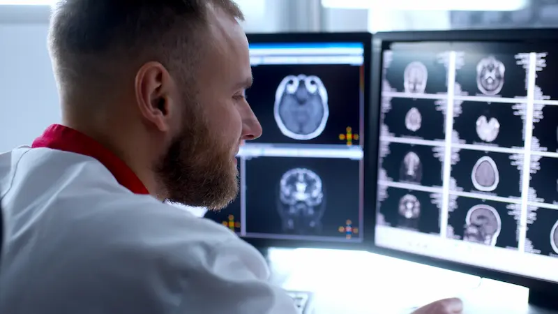
When the brain MRI procedure is finished, the technologist will slide the table out of the scanner and remove any coils or IV lines. You can immediately sit up and get dressed. Most people feel completely normal afterward and can resume their daily activities right away.
Resuming Your Normal Activities Immediately
If your exam was done without sedation or contrast, you can eat, drink, and return to work or driving right after. The magnetic field does not stay in your body, so there are no lasting sensations or restrictions.
Special Instructions (If You Had Sedation or Contrast)
Patients who received contrast dye should drink plenty of water afterward to help flush the material from their system. If sedation was used, rest for the remainder of the day and avoid driving or operating heavy machinery until the medication wears off.
Although side effects from contrast dye are rare, contact your doctor if you notice a rash, swelling, or persistent nausea. These are uncommon but manageable allergic reactions.
How and When Will You Get Your Results?
A radiologist will review your images, analyze every detail, and prepare a report for your referring physician. In most clinics, results are available within 24 to 48 hours. Your doctor will discuss the findings and explain what they mean in relation to your symptoms.
If abnormalities are detected, additional tests may be recommended. However, many brain MRI scans show normal results, which helps doctors rule out serious conditions.
Frequently Asked Questions (FAQ)
How long does a brain MRI take?
A brain MRI usually takes 30–45 minutes. If contrast dye is needed, it may last up to an hour. Staying still during the scan ensures clear results.
Is a brain MRI painful?
No. The brain MRI procedure is painless. You may hear loud noises and feel a slight pinch if contrast is used, but there is no actual discomfort.
What can a brain MRI detect?
A head MRI can reveal tumors, strokes, infections, multiple sclerosis, or inflammation. It helps doctors see brain structures and diagnose neurological issues.
What to wear for an MRI?
Wear loose, metal-free clothing or change into a gown. Remove jewelry, watches, and hair accessories to avoid interference with the magnetic field.