
Welcome! If you’re exploring options for medical imaging abroad, you’ve likely discovered that Turkey is a leading destination for high-quality, accessible healthcare. This comprehensive guide is specifically designed for international patients like you who are considering or planning to have an ultrasound in Turkey. We understand that navigating healthcare in another country can seem daunting, so we’ve gathered essential information all in one place. Here, you’ll learn about the technology itself, common ultrasound procedures performed, the significant advantages of choosing Turkey’s top clinics and specialists, cost comparisons, how to prepare for your examination, and answers to frequently asked questions. Our goal is to provide clarity and confidence as you explore your options for ultrasound in Turkey.
Table of Contents
Understanding Ultrasound Technology
Medical imaging plays a crucial role in modern healthcare, allowing doctors to see inside the human body without the need for surgery. Among the various imaging techniques available, ultrasound stands out for its safety, versatility, and effectiveness. For international patients considering medical procedures abroad, understanding the technology behind an ultrasound in Turkey is the first step towards making an informed decision. This guide will delve into what ultrasound is, how it works, its history, and its key advantages, particularly for those seeking high-quality diagnostic services in world-class Turkish facilities.
Choosing to have an ultrasound in Turkey means accessing advanced diagnostic capabilities in a country renowned for its medical expertise and patient-centred care. Whether for routine check-ups, pregnancy monitoring, or investigating specific symptoms, ultrasound provides valuable insights quickly and safely. Let’s explore the fundamentals of this remarkable technology.
What Is Ultrasound and How Do Sound Waves Create an Image?
Key takeaway: Ultrasound, also known professionally as sonography, is a medical imaging technique that uses high-frequency sound waves—far above the range of human hearing—to create real-time pictures of the inside of your body. It does not use any ionising radiation (like X-rays), making it exceptionally safe for all patients, including pregnant women and children considering an ultrasound in Turkey.
The process of creating an ultrasound image begins with a skilled ultrasound tech, often called a sonographer. They use a small, hand-held device called a transducer, or probe. This device does two things: it sends pulses of high-frequency sound waves into the body and then listens for the echoes that bounce back. To ensure the sound waves travel efficiently from the transducer into the body and aren’t blocked by air, a special water-based gel is applied to the skin over the area being examined. This might feel a little cold initially, but it’s harmless. This is a standard part of getting an ultrasound in Turkey.
As the sound waves travel through the body, they encounter different types of tissues, such as organs, fluids, and bones. Each type of tissue reflects the sound waves differently. For example, dense structures like bone reflect a lot of sound waves, while fluid-filled areas like cysts let most waves pass through, reflecting very little. The sound waves that bounce back (the echoes) are detected by the same transducer that sent them out. The strength of these returning echoes and the time it takes for them to come back provide crucial information. An ultrasound in Turkey uses this principle effectively.
A powerful computer connected to the transducer processes these returning echoes instantly. It calculates how far the wave travelled based on the time delay and how strong the echo was. Using this data, it constructs a live, moving image on a monitor. This generated picture is technically called a sonogram, though the terms ultrasound and sonogram are often used interchangeably. Different tissues appear as varying shades of grey on the sonogram: fluid typically looks black because it sends back few echoes, while solid tissues or structures appear lighter grey or white. Getting an ultrasound in Turkey means benefiting from this sophisticated image generation.
One of the unique strengths of ultrasound imaging is its ability to show movement in real-time. Doctors can watch organs function, observe blood flow through vessels (using a special type called Doppler ultrasound), or see a baby move during a pregnancy scan. After the sonographer captures the necessary images during your ultrasound in Turkey, a specialist doctor, usually a radiologist or another physician trained in interpreting these scans, will analyse the sonogram images to make a diagnosis or assessment. The quality of your ultrasound in Turkey depends significantly on both the technology and the expertise of the ultrasound doctor and ultrasound tech.
From Discovery to Today: A Brief History of Medical Ultrasound
Key takeaway: The journey of medical ultrasound from its conceptual origins to the sophisticated diagnostic tool used today, including for ultrasound in Turkey, is a fascinating story of scientific innovation spanning several decades. Its roots lie not in medicine, but in understanding sound wave behavior and naval technology.
The underlying principle of using sound waves to detect objects dates back to the late 18th century with studies on bat navigation and early experiments measuring the speed of sound in water. However, the practical application began gathering pace in the early 20th century. During World War I, physicist Paul Langevin developed “sonar” (SOund Navigation And Ranging) using high-frequency sound waves (ultrasound) to detect submerged submarines. This foundational work laid the groundwork for future medical applications. Having an ultrasound in Turkey connects you to this lineage of innovation.
The first documented attempt to use ultrasound for medical diagnosis was in the 1940s by Austrian neurologist Dr. Karl Dussik, who tried to image the brain’s ventricles to detect tumours. While his initial methods were basic and faced limitations (sound waves don’t travel well through bone), it marked the birth of diagnostic medical ultrasound. The true potential began to unfold in the 1950s.
Quick list: Key Milestones
- 1940s: Dr. Karl Dussik attempts brain imaging.
- 1950s: Prof. Ian Donald pioneers obstetric ultrasound. A-mode and early B-mode scanners developed.
- 1960s-70s: Applications expand to abdomen, heart (echocardiography), and blood vessels. Grayscale imaging improves image quality.
- 1980s-90s: Digital revolution brings higher resolution, portability, and Doppler ultrasound for blood flow assessment. This era influences the technology used for ultrasound in Turkey today.
- 2000s-Present: Real-time 3D and 4D imaging become widespread, especially in obstetrics. Miniaturisation leads to portable and point-of-care devices. The standard for an ultrasound in Turkey includes these advanced features.
A major breakthrough came in the 1950s thanks to Scottish obstetrician Professor Ian Donald and engineer Tom Brown. Working with industrial flaw detectors, they adapted the technology for medical use. In 1958, Donald published landmark papers demonstrating the use of ultrasound to examine foetuses and differentiate between various gynaecological conditions, like cysts and tumours. His work established ultrasound as a vital tool in obstetrics and gynaecology, earning him the title “father of obstetric ultrasound.” Early machines produced simple one-dimensional (A-mode) or basic two-dimensional (B-mode) static images. Modern ultrasound procedures build upon this foundation.
Throughout the 1960s and 1970s, ultrasound technology rapidly evolved. Researchers explored its use in imaging abdominal organs, the heart (echocardiography), and blood vessels. The introduction of grayscale imaging allowed for much more detailed visualisation of tissue textures, enhancing diagnostic accuracy significantly. This period saw ultrasound transition from an experimental technique to a mainstream clinical tool, setting the stage for the advanced ultrasound in Turkey available now.
The digital age of the 1980s and 1990s brought transformative changes. Computer processing power led to vastly improved image resolution, smaller and more portable machines, and new functional capabilities. Doppler ultrasound, which allows visualisation and measurement of blood flow, became a critical tool in cardiology and vascular studies. Later, the development of real-time 3D and 4D ultrasound provided stunningly detailed, volumetric views, particularly popular for viewing foetuses during pregnancy. Today’s ultrasound in Turkey utilises this cutting-edge technology.
Key Advantages: Non-Invasive, Real-Time, and Radiation-Free
Key takeaway: Ultrasound imaging offers compelling advantages that make it a preferred choice for many diagnostic situations, particularly for patients seeking safe and effective procedures like ultrasound in Turkey. Its three primary benefits are that it is non-invasive, provides real-time imaging, and is completely free from ionizing radiation.
- Non-Invasive: Perhaps the most significant advantage for patients is that standard external ultrasound is non-invasive. This means no needles, injections, cuts, or entry into the body is typically required. The ultrasound tech simply places the transducer coated with gel onto your skin and moves it gently over the area of interest. This lack of invasiveness translates to a generally painless and comfortable experience, a major plus when considering an ultrasound in Turkey. Some specialized ultrasound procedures might involve internal probes (like transvaginal or transrectal scans), but even these are minimally invasive compared to surgery.
- Real-Time Imaging: Unlike static images produced by X-rays or some MRI sequences, ultrasound provides dynamic, real-time visualization. The ultrasound doctor or tech can see structures inside the body moving as they actually happen. This includes observing a baby’s movements and heartbeat during pregnancy, watching heart valves function, assessing blood flow through arteries and veins, or guiding procedures like biopsies with precision. This ‘live’ view is invaluable for accurate diagnosis and assessment, a key feature of ultrasound in Turkey services.
- Radiation-Free: This is a critical safety feature. Ultrasound imaging uses high-frequency sound waves, not ionizing radiation like X-rays or CT scans. Ionizing radiation, especially in cumulative doses, carries potential long-term risks. Because ultrasound avoids this radiation entirely, it is considered exceptionally safe. There are no known harmful side effects associated with diagnostic ultrasound when used correctly. This makes it the ideal imaging modality for sensitive situations, such as monitoring foetal development throughout pregnancy and examining children. Patients choosing ultrasound in Turkey can be assured of this safety standard.
Quick list: Comparing Ultrasound Advantages
- Ultrasound vs. X-ray: Ultrasound shows soft tissues better; X-ray shows bones better. Ultrasound is radiation-free; X-ray uses ionising radiation.
- Ultrasound vs. CT Scan: Ultrasound is radiation-free; CT uses significant ionizing radiation. Ultrasound is generally less expensive and more portable. CT provides highly detailed cross-sectional images, sometimes better for bone or complex issues.
- Ultrasound vs. MRI: Both are radiation-free. MRI often provides superior soft tissue detail, especially for the brain and joints. However, MRI is much more expensive, takes longer, can be noisy and confining, and is not suitable for patients with certain metal implants. Ultrasound is faster, cheaper, widely available, and excellent for many applications, including real-time imaging, making ultrasound in Turkey a practical choice.
Beyond these core advantages, ultrasound is also widely accessible, relatively inexpensive compared to CT or MRI, and highly portable, with some machines being laptop-sized or even hand-held. Its ability to clearly visualize soft tissues, fluids, and blood flow makes it indispensable across numerous medical specialties. These combined benefits solidify the value of choosing ultrasound in Turkey for your diagnostic needs. The technology provides powerful insights without compromising patient safety or comfort.
Common Ultrasound Procedures Performed in Turkey
Key takeaway: Turkey’s modern medical facilities offer a comprehensive array of ultrasound procedures, utilising advanced technology to provide accurate diagnoses for a wide range of conditions. From routine check-ups to specialized investigations, you can confidently access the specific type of ultrasound in Turkey you need. Understanding these common procedures can help you know what to expect and the valuable information these scans provide.
Ultrasound imaging is incredibly versatile. Depending on the part of the body being examined and the clinical question being asked, different techniques and types of ultrasound probes may be used. Clinics providing ultrasound in Turkey are well-equipped to perform standard examinations as well as more complex ultrasound procedures, ensuring patients receive appropriate and thorough diagnostic care delivered by experienced professionals. Let’s explore some of the most frequently performed ultrasound examinations.
Abdominal & Hepatobiliary Ultrasound
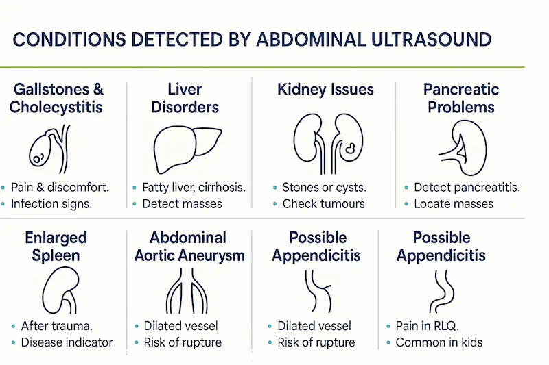
Key takeaway: An abdominal ultrasound provides detailed images of the major organs within your abdominal cavity. This includes the liver, gallbladder, spleen, pancreas, kidneys, and abdominal aorta. It is a cornerstone diagnostic tool for investigating abdominal pain, abnormal blood tests, or suspected organ issues, and is commonly performed as part of health screenings or specific diagnostic pathways for ultrasound in Turkey.
This type of ultrasound is essential for detecting a variety of conditions. For instance, it’s excellent for finding gallstones within the gallbladder or bile ducts, identifying liver problems like fatty liver disease, cirrhosis, cysts, or tumors, and assessing the kidneys for stones, blockages (hydro nephrosis), cysts, or tumors. The pancreas and spleen are also evaluated for abnormalities. An abdominal ultrasound in Turkey can also be used to screen for an abdominal aortic aneurysm (AAA), a potentially dangerous swelling in the main artery supplying blood to the lower body.
Quick list: Conditions Detected by Abdominal Ultrasound
- Gallstones and gallbladder inflammation (cholecystitis)
- Liver conditions (fatty liver, cirrhosis, cysts, masses)
- Kidney stones, blockages, cysts, and tumours
- Pancreatitis or pancreatic masses
- Spleen enlargement or damage
- Abdominal Aortic Aneurysm (AAA)
- Sometimes, signs of appendicitis (especially in children)
Typically, an abdominal ultrasound requires some preparation. You’ll usually be asked to fast (not eat or drink anything except water) for several hours before the scan. This helps in two ways: it ensures your gallbladder is full and easier to see, and it reduces the amount of gas in your intestines, which can interfere with the sound waves and obscure the image. During the procedure, you’ll lie comfortably while the ultrasound tech applies gel and moves the transducer across your abdomen. Having this type of ultrasound in Turkey is a straightforward and painless process.
Pelvic and Transvaginal Ultrasound
Key takeaway: Pelvic ultrasound focuses on the organs within the pelvic region. In women, this primarily includes the uterus, ovaries, fallopian tubes, cervix, and bladder. In men, it can assess the bladder and prostate gland. There are two main approaches: transabdominal (through the abdominal wall) and transvaginal (using a specialized internal probe), both readily available for ultrasound in Turkey depending on the diagnostic need.
A transabdominal pelvic ultrasound is performed similarly to an abdominal scan, with the transducer moved across the lower abdomen. For this approach, you might be asked to drink water beforehand to ensure your bladder is full, which acts as an acoustic ‘window’, pushing the bowel out of the way and helping to provide clearer images of the pelvic organs. This method gives a good overview of the pelvic structures. Getting this ultrasound in Turkey is common for initial assessments.
For a more detailed view, especially of the uterus lining (endometrium), ovaries, and early pregnancy, a transvaginal ultrasound is often preferred. For this procedure, a thin, specially designed transducer probe, covered with a protective sheath and lubricant, is gently inserted into the vagina. While this might sound uncomfortable, it is generally well-tolerated and provides much clearer, higher-resolution images because the probe is closer to the pelvic organs. Clinics offering ultrasound in Turkey ensure patient comfort and privacy during this procedure.
Quick list: Conditions Assessed by Pelvic Ultrasound
- Uterine fibroids or polyps
- Ovarian cysts or tumors
- Endometriosis assessment (indirect signs)
- Pelvic Inflammatory Disease (PID)
- Ectopic pregnancy (pregnancy outside the uterus)
- Causes of abnormal bleeding or pelvic pain
- Infertility investigations (e.g., monitoring follicles)
- Checking Intrauterine Device (IUD) position
- Bladder issues (volume, wall thickness)
- Prostate assessment (often combined with transrectal ultrasound in men)
The choice between transabdominal and transvaginal ultrasound depends on the specific reason for the scan. Often, both may be performed during the same appointment to get a complete picture. Your doctor or the clinic performing your ultrasound in Turkey will explain which approach is best for you.
Obstetric & Fetal Sonography
Key takeaway: Obstetric ultrasound, or fetal sonography, is the use of ultrasound to monitor the health and development of a baby during pregnancy. It’s a safe and invaluable tool used routinely worldwide, allowing parents and doctors to visualize the fetus, confirm viability, estimate the due date, assess growth, and screen for potential abnormalities. Facilities offering ultrasound in Turkey provide comprehensive obstetric scanning services, including advanced 3D and 4D imaging.
Ultrasound plays a critical role throughout pregnancy. Early scans (first trimester) confirm the pregnancy is located within the uterus, detect the fetal heartbeat, determine the number of fetuses, and accurately estimate the gestational age and due date. A key scan around 11-14 weeks, often combined with blood tests, screens for chromosomal abnormalities like Down syndrome by measuring the nuchal translucency (fluid at the back of the baby’s neck). Accessing early ultrasound in Turkey can provide crucial reassurance.
Around 18-20 weeks (second trimester), a detailed anatomy scan is typically performed. This involves a thorough examination of the baby’s major organs, limbs, face, and overall structure to check for normal development and detect any potential congenital anomalies. The scan also assesses fetal growth, amniotic fluid levels, and the position of the placenta. Later in pregnancy (third trimester), ultrasound may be used to monitor growth, check the baby’s position before delivery, assess placental function, or evaluate fetal well-being if concerns arise. An ultrasound in Turkey ensures these vital checks are performed to high standards.
Modern ultrasound technology now includes 3D and 4D capabilities, which are particularly popular in obstetrics. 3D ultrasound compiles the sound wave data to create still, three-dimensional images of the baby, offering a clearer view of surface features like the face. 4D ultrasound adds the dimension of time, creating moving 3D images – essentially a live video. While not always medically necessary, these scans can enhance parental bonding and sometimes help in visualizing certain abnormalities more clearly. Many clinics providing ultrasound in Turkey offer these advanced imaging options.
Cardiac (Echocardiography) Ultrasound
Key takeaway: An echocardiogram, often called an “echo,” is a specialized ultrasound examination focused entirely on the heart. It provides detailed images of the heart’s chambers, valves, walls, and the surrounding blood vessels (like the aorta), allowing doctors to assess its structure and function non-invasively. High-quality cardiac ultrasound in Turkey is performed by trained specialists in well-equipped cardiology departments or clinics.
An echocardiogram is crucial for diagnosing and managing a wide range of heart conditions. It allows doctors to evaluate the size and thickness of the heart chambers, assess how well the heart muscle is contracting and relaxing (its pumping function, often measured as ejection fraction), and examine the structure and movement of the heart valves. Valve problems like stenosis (narrowing) or regurgitation (leaking) can be clearly identified. Getting an ultrasound in Turkey for cardiac assessment is common.
Quick list: What Echocardiography Assesses
- Heart chamber size and wall thickness
- Pumping strength (ejection fraction)
- Heart valve structure and function (stenosis, regurgitation)
- Presence of blood clots within the heart
- Detection of tumors or abnormal growths
- Assessment of congenital heart defects
- Evaluation of fluid around the heart (pericardial effusion)
- Monitoring changes after heart attack or surgery
The most common type is a transthoracic echocardiogram (TTE), where the transducer is moved across the chest wall. Gel is applied, and you may be asked to lie in different positions or hold your breath briefly to get the best images. In some cases, if clearer pictures are needed (especially of structures at the back of the heart), a transesophageal echocardiogram (TEE) might be performed. This involves passing a small probe down the throat into the esophagus, which sits directly behind the heart. A stress echocardiogram involves performing an echo before and immediately after exercise (usually on a treadmill) to see how the heart functions under stress. Clinics performing cardiac ultrasound in Turkey offer these variations.
Vascular, Doppler, and Blood-Flow Studies
Key takeaway: Vascular ultrasound uses Doppler technology to specifically examine blood flow within arteries and veins throughout the body. This is vital for detecting blockages, narrowing, blood clots (like DVT), aneurysms (bulges in vessel walls), and other vascular abnormalities. Timely vascular ultrasound in Turkey can help diagnose conditions that could lead to serious complications like stroke or pulmonary embolism.
Doppler ultrasound works by measuring the frequency shift of sound waves as they reflect off moving red blood cells. This allows the ultrasound machine to calculate the speed and direction of blood flow. Often, this information is displayed visually using colour (Colour Doppler), where different colours (typically red and blue) represent flow towards or away from the transducer. This provides an intuitive map of blood circulation within the vessels being examined. This technology is standard for vascular ultrasound in Turkey.
Common applications for vascular and Doppler ultrasound include:
- Carotid Ultrasound: Examines the carotid arteries in the neck, which supply blood to the brain. It looks for plaque buildup (atherosclerosis) that can narrow the arteries and increase stroke risk.
- Lower Limb Venous Ultrasound: Checks the deep veins in the legs for blood clots (Deep Vein Thrombosis – DVT), which can be life-threatening if they travel to the lungs (pulmonary embolism).
- Lower Limb Arterial Ultrasound: Assesses blood flow in the leg arteries to diagnose Peripheral Artery Disease (PAD), which causes pain and cramping during walking (claudication) due to poor circulation.
- Renal Ultrasound with Doppler: Evaluates blood flow to and from the kidneys, often used to investigate high blood pressure.
- Abdominal Doppler: Checks blood flow in major abdominal vessels like the aorta, vena cava, and arteries supplying the liver or intestines.
Like other ultrasound procedures, vascular studies are typically non-invasive and painless, involving gel and a transducer moved over the skin. They provide critical information about circulatory health without risks or discomfort. Opting for a vascular ultrasound in Turkey gives you access to essential preventive and diagnostic screening using sophisticated Doppler techniques.
Why Choose Ultrasound Imaging in Turkey?
Key takeaway: Turkey has rapidly emerged as a global hub for medical tourism, and for good reason. Choosing to have your ultrasound imaging performed here offers a compelling blend of high-quality medical care, access to advanced technology, experienced professionals, and significant cost advantages, all within a patient-friendly environment. Opting for an ultrasound in Turkey is not just about affordability; it’s about receiving world-class diagnostic services you can trust.
Beyond the technical capabilities of ultrasound explored earlier, several factors make Turkey an attractive destination for international patients seeking diagnostic tests. The healthcare system has invested heavily in infrastructure and personnel, creating centers of excellence that rival those in Western Europe and North America. When considering an ultrasound in Turkey, you are choosing a pathway that prioritizes both clinical excellence and patient experience, often with shorter waiting times than you might encounter back home.
Best Clinics and Centrally Located Imaging Centers
Key takeaway: Turkey is home to numerous modern hospitals and specialised diagnostic centres equipped with the latest ultrasound technology. Many of these facilities, particularly the best clinic for ultrasound in Turkey options found in major hubs like Istanbul, Ankara, Antalya, and İzmir, hold prestigious international accreditations, signifying their commitment to global standards of quality and patient safety.
One of the primary indicators of quality is accreditation by bodies such as the Joint Commission International (JCI). Numerous Turkish hospitals and clinics have achieved JCI accreditation, demonstrating that their facilities, procedures, and patient safety protocols meet rigorous international benchmarks. This provides significant reassurance for patients travelling for an ultrasound in Turkey. These accredited centers invest heavily in maintaining state-of-the-art diagnostic equipment, ensuring your ultrasound imaging is performed using machines with high resolution, advanced Doppler capabilities for blood flow assessment, and often 3D/4D sonography options where relevant (like in obstetric ultrasound).
Finding the best clinic for ultrasound in Turkey also involves considering convenience. Major healthcare providers often have multiple branches, strategically located within large cities. These centers are typically easily accessible via public transport or taxi and are often situated relatively close to international airports and a range of accommodation options, simplifying the logistics for overseas visitors. Whether you need a routine check or a complex diagnostic ultrasound procedure, you’ll find well-equipped, easily reachable facilities ready to assist you. The infrastructure supporting medical tourism for services like ultrasound in Turkey is well-developed.
Furthermore, these clinics are designed with patient comfort in mind, featuring modern amenities, clean environments, and efficient workflows to minimize waiting times. From the moment you inquire about an ultrasound in Turkey to receiving your results, reputable clinics strive to provide a seamless and positive experience, understanding the unique needs of international patients. They offer a level of service and technological advancement that meets or exceeds expectations.
Internationally Acclaimed Doctors & Certified Sonography Techs
Key takeaway: The quality of any medical procedure, including an ultrasound, heavily relies on the expertise of the professionals involved. Turkey boasts a pool of highly qualified, often internationally trained radiologists, specialist physicians, and certified ultrasound technologists (sonographers) who ensure that your ultrasound in Turkey is performed and interpreted accurately.
The ultrasound doctor responsible for interpreting your scan images is typically a board-certified radiologist or a specialist relevant to the examination (e.g., a cardiologist for an echocardiogram, an obstetrician-gynecologists for fetal or pelvic scans). Many of these doctors have gained experience or further training in leading medical institutions across Europe or the United States, bringing a wealth of international knowledge to their practice in Turkey. Finding the best doctor for ultrasound in Turkey often means looking for professionals affiliated with major, accredited hospitals known for their expertise in specific fields. Their credentials often include memberships in respected international medical societies and publications in peer-reviewed journals, reflecting their commitment to their field.
Equally important is the skill of the ultrasound tech or sonographer who performs the actual scan. Obtaining clear, diagnostic-quality ultrasound images requires technical proficiency, a good understanding of anatomy, and careful attention to detail. Sonographers working in reputable centers providing ultrasound in Turkey are typically certified professionals with specialized training. They know how to operate the sophisticated equipment effectively, optimize the images, and ensure all necessary views are captured for the interpreting ultrasound doctor. Their expertise is fundamental to the accuracy of your ultrasound imaging.
Communication is also key for international patients. Recognising this, many leading hospitals and clinics offering ultrasound in Turkey employ multilingual staff, particularly doctors and patient coordinators who speak fluent English. Translation services are also commonly available if needed, ensuring you can comfortably discuss your concerns, understand the ultrasound procedure, and receive your results clearly. This focus on communication enhances the overall experience and contributes to the trustworthiness of ultrasound in Turkey.
Patient Reviews, Success Stories, and After-Care Support
Key takeaway: Choosing to have a medical procedure abroad involves trust. Positive patient experiences, readily available testimonials, and comprehensive support services offered by Turkish clinics provide strong indicators of reliability and patient satisfaction for those considering an ultrasound in Turkey. The focus extends beyond the scan itself to ensure a smooth overall journey.
Many international patients share their experiences online through reviews and testimonials. Reading about the journeys of others who have had an ultrasound in Turkey can offer valuable insights into the quality of care, the professionalism of the staff, the efficiency of the service, and the overall patient experience at specific clinics. While individual experiences vary, a consistent pattern of positive feedback is a strong sign of a reputable provider. These firsthand accounts often highlight the welcoming atmosphere and attentive care received during their ultrasound procedures.
A “success story” in the context of diagnostic ultrasound imaging means receiving a timely, accurate diagnosis that enables appropriate next steps. Whether it’s the reassurance of a healthy pregnancy sonogram, the early detection of a condition allowing for prompt treatment, or clear images guiding a subsequent medical intervention, the effectiveness of the ultrasound in Turkey is paramount. Clinics pride themselves on contributing to positive health outcomes through high-quality diagnostics. The detailed image quality from modern ultrasound machines plays a big role here.
Beyond the clinical aspects, leading centers providing ultrasound in Turkey understand the needs of medical tourists and offer robust support systems. This often includes:
- Appointment Coordination: Assistance with scheduling your ultrasound and any related consultations.
- Language Services: Providing interpreters or ensuring communication with English-speaking staff.
- Logistical Help: Offering guidance or packages that may include airport transfers and accommodation suggestions.
- Clear Communication: Ensuring you understand the preparation required, the procedure itself, and how and when you will receive your results.
- Results Delivery: Providing comprehensive reports (often in English) and digital copies of your ultrasound images promptly, making it easy to share with your doctor back home.
- After-Care Guidance: Explaining the findings and advising on necessary follow-up actions or connecting you with specialists if needed.
This comprehensive support network makes the process of getting an ultrasound in Turkey significantly less stressful, allowing patients to focus on their health. The commitment to patient well-being from start to finish is a hallmark of the top healthcare providers in the country.
Cost of Ultrasound in Turkey
Key takeaway: One of the most significant advantages drawing international patients to Turkey for medical services is the exceptional cost-effectiveness, and this certainly applies to diagnostic procedures like ultrasound. The cost of ultrasound in Turkey is considerably lower than in many Western countries, yet this affordability does not come at the expense of quality. Patients receive high-standard ultrasound imaging using modern technology, interpreted by skilled professionals, all at a fraction of the price they might expect to pay at home.
Understanding the potential expenses involved is crucial when planning medical travel. While exact prices can vary, Turkey consistently offers highly competitive rates for a wide range of ultrasound procedures. This section will provide insights into typical price ranges across major Turkish cities, compare these costs to those in Europe and the US, and outline the key factors that influence the final cost of ultrasound in Turkey, helping you budget effectively for your diagnostic needs as of May 2025. Getting an ultrasound in Turkey represents excellent value for money.
Price Comparison: Istanbul, Ankara, Antalya, and İzmir
Key takeaway: While the overall cost of ultrasound in Turkey is favorable across the board, minor variations can exist between major cities known for medical tourism. Generally, prices in large metropolitan hubs like Istanbul and the capital, Ankara, might be slightly higher than in coastal cities like Antalya or İzmir, but the difference is often not substantial, and affordability remains a key feature everywhere. High-quality ultrasound in Turkey is accessible nationwide.
Based on current estimates (as of May 2025), the price for common ultrasound examinations in Turkey can vary. For example:
- Abdominal Ultrasound: Prices might range roughly from $50 to $250 USD (approximately €45 – €230 EUR).
- Pelvic Ultrasound (Transabdominal/Transvaginal): Similar ranges often apply, perhaps $60 – $300 USD (approximately €55 – €275 EUR).
- Obstetric Ultrasound (Standard): Costs could fall between $70 – $350 USD (approximately €65 – €320 EUR), potentially higher for detailed anomaly scans or 3D/4D options.
- Thyroid or Breast Ultrasound: Often priced similarly to abdominal or pelvic scans, perhaps in the $50 – $250 USD range.
- Doppler Ultrasound (e.g., Carotid, Limb): May range from $100 – $400 USD (approximately €90 – €370 EUR) due to the specialised technology.
- Echocardiogram (Cardiac Ultrasound): Typically ranges from $150 – $500 USD (approximately €140 – €460 EUR), depending on complexity.
Important Note: These figures are estimates and can fluctuate based on the specific clinic, required detail, and included services. Always request a precise quote from your chosen provider for your ultrasound in Turkey. Some sources report average costs around $220 USD (€200 EUR), but individual procedure costs vary widely.
Why the slight city variations? Factors include the general cost of living and operating businesses, the level of competition among healthcare providers, and the specific reputation or ‘brand value’ of certain clinics, which may be more concentrated in Istanbul or Ankara. However, excellent facilities providing top-notch ultrasound in Turkey exist in all these major cities. Antalya and İzmir, being popular tourist destinations, also have strong healthcare infrastructures catering to international visitors, often combining high quality with potentially more competitive pricing for ultrasound procedures.
Additionally, many clinics specializing in medical tourism offer package deals. These might bundle the cost of ultrasound in Turkey with consultations, translation services, or even airport transfers. While potentially convenient, it’s wise to understand what’s included in any package price to make accurate comparisons.
Turkey vs. Europe & the U.S.: How Much Can You Save?
Key takeaway: The difference in the cost of ultrasound in Turkey compared to Western Europe and especially the United States is often dramatic. Patients can realistically expect to save anywhere from 50% to 80%, and sometimes even more, on the cost of ultrasound procedures without compromising the quality of care, technology, or expertise.
Let’s look at some broad comparisons (remembering that prices abroad vary greatly depending on the country, insurance status, and whether care is public or private):
- United States: The cost of an ultrasound can range widely, often from $300 USD for a simple scan in an outpatient setting to well over $1,000 USD or even $2,000+ USD in a hospital setting, particularly for more complex studies like detailed echocardiograms or obstetric scans. Insurance negotiations significantly impact final patient costs, but the ‘list price’ is often very high.
- Western Europe (e.g., UK, Germany, France): In private settings, ultrasound costs might typically range from €200 EUR to €800 EUR ($220 – $880 USD approximately) or more, depending on the scan type and country. While public healthcare systems may cover costs for residents, waiting times can be long, and non-residents or those seeking elective scans often face significant private fees.
Comparing these figures with the estimated ranges for ultrasound in Turkey (e.g., $50 – $500 USD depending on type), the potential savings are clear and substantial. A procedure costing $800 USD in the US might be available for $200 USD in Turkey – a 75% saving. This affordability makes essential diagnostic ultrasound imaging much more accessible, especially for those facing high deductibles, long waiting lists, or lack of insurance coverage at home.
This remarkable value proposition is a key driver for medical tourism to Turkey. The savings achieved on the cost of ultrasound in Turkey can often offset travel expenses, and patients can access necessary diagnostics quickly and efficiently. It allows individuals to take proactive steps for their health without facing prohibitive costs. Clinics offering ultrasound in Turkey are accustomed to providing clear price information to international patients, enhancing transparency.
Factors That Affect Ultrasound Pricing (Clinic, Doctor, Equipment)
Key takeaway: While the overall cost of ultrasound in Turkey is low, several factors contribute to the specific price quoted by a clinic or hospital. Understanding these elements helps explain potential variations and allows patients to assess the value offered by different providers for their ultrasound in Turkey.
- Clinic Reputation and Accreditation: Highly reputable hospitals or clinics, especially those with international accreditation (like JCI) and a strong brand presence, may charge a premium. This often reflects investment in top-tier facilities, comprehensive patient services, robust quality control, and potentially higher administrative overheads. Their ultrasound price reflects this overall package.
- Doctor’s Experience and Specialization: The expertise of the interpreting ultrasound doctor can influence cost. Scans interpreted by a renowned professor, a sub-specialist (e.g., a pediatric cardiologist for a fetal echo), or a doctor with extensive international experience might command a higher fee than a standard interpretation by a general radiologist. The cost of ultrasound in Turkey can reflect this level of expertise.
- Type and Complexity of the Ultrasound: This is a major factor. A basic, quick scan of a single area (like the thyroid) will naturally cost less than a comprehensive abdominal ultrasound, a detailed fetal anomaly scan requiring an hour or more, or a specialized study like a transesophageal echocardiogram or a contrast-enhanced ultrasound. Procedures requiring more time, advanced techniques (like 3D/4D or specific Doppler measurements), or specialized probes will have a higher cost of ultrasound in Turkey.
- Technology and Equipment: While most reputable centers use modern equipment, clinics equipped with the very latest, cutting-edge ultrasound machines might incorporate this technological advantage into their pricing structure. Features like premium image resolution or advanced software capabilities can impact the ultrasound procedure cost.
- Geographic Location within Turkey: As mentioned earlier, operating costs vary between cities and even districts within a city. Clinics in prime locations in Istanbul or Ankara might have higher rents and operational expenses, which can be reflected subtly in the cost of ultrasound in Turkey.
- Package Inclusions: If the quoted price is part of a package, ensure you know what it covers. Does it include just the scan and report, or also a pre-scan consultation, post-scan discussion with the ultrasound doctor, translation services, or other amenities? This affects the perceived cost of ultrasound in Turkey.
By considering these factors, patients can better understand the pricing structure for ultrasound in Turkey and choose a provider that offers the best balance of cost, quality, expertise, and convenience for their specific needs. Always aim for clear, itemised quotes.

Preparing for Your Ultrasound Examination
Key takeaway: While ultrasound is generally a straightforward and non-invasive procedure, some preparation may be required depending on the specific type of scan you are having. Following the preparation instructions provided by the clinic performing your ultrasound in Turkey is essential for obtaining the clearest possible images and ensuring an accurate diagnosis.
Preparation steps are designed to optimise the conditions for ultrasound imaging. For instance, reducing gas in the bowel or ensuring the bladder is full can significantly improve the quality of the sonogram. The staff at your chosen facility for ultrasound in Turkey are experienced in guiding international patients and will provide clear, specific instructions beforehand, often in English or via translation services. Don’t hesitate to ask questions if anything is unclear.
Pre-Scan Checklist: Fasting, Hydration, Clothing, Medications
Key takeaway: Preparation requirements vary widely depending on the area being scanned. Common instructions may involve fasting (not eating), specific hydration (drinking water to fill the bladder), wearing appropriate clothing, and informing the clinic about your current medications. Adhering to these guidelines before your ultrasound in Turkey is vital for a successful examination.
Here’s a general checklist, but always follow the specific advice given to you:
- Fasting: For ultrasound examinations of the abdomen (especially focusing on the gallbladder, liver, or pancreas), you will likely be asked to fast – meaning no food or drink, except sips of water – for about 6 to 8 hours before your appointment. Fasting prevents the gallbladder from contracting after eating, making it easier to visualize, and also helps reduce gas in the intestines which can block the sound waves.
- Hydration / Full Bladder: For transabdominal pelvic ultrasound scans (looking at the uterus, ovaries, or bladder itself), you’ll typically be instructed to drink a significant amount of water (often around 1 liter or 4-5 glasses) about one hour before the scan and not empty your bladder. A full bladder pushes the bowel loops aside and provides a clear ‘window’ for the sound waves to reach the pelvic organs. This is crucial for getting good quality images during this type of ultrasound in Turkey. For other scans, like vascular or thyroid ultrasound, this is usually not necessary.
- Clothing: Wear loose-fitting, comfortable clothing that allows easy access to the part of your body being examined. You might be asked to remove some clothing and wear a hospital gown, depending on the area being scanned. Avoid wearing jewelry in the examination area.
- Medications: Generally, you can continue taking your prescribed medications as usual unless specifically told otherwise by your doctor or the ultrasound clinic. However, it’s crucial to inform the clinic performing your ultrasound in Turkey about all medications and supplements you are taking when you book your appointment.
- Lotions and Powders: Avoid applying lotions, creams, or powders to the skin in the area to be scanned on the day of your ultrasound procedure, as these can sometimes interfere with the transducer contact.
Always confirm the specific preparation instructions for your particular ultrasound in Turkey well in advance.
Step-by-Step: What to Expect During the Procedure
Key takeaway: Knowing what typically happens during an ultrasound scan can help alleviate any anxiety. The process is usually quick, painless, and involves lying comfortably while a trained ultrasound tech (sonographer) moves a probe over your skin using a special gel. Clinics providing ultrasound in Turkey prioritize patient comfort and clear communication throughout the procedure.
Here’s a typical step-by-step guide for an external ultrasound:
- Step 1: Arrival & Check-in: You’ll arrive at the clinic or hospital department, check in for your appointment, and may be asked to wait briefly. Depending on the scan, you might be asked to change into a gown.
- Step 2: Positioning: The sonographer will escort you to the examination room and ask you to lie down on a padded table. Depending on the area being scanned, you might lie on your back, side, or occasionally your front.
- Step 3: Gel Application: The ultrasound tech will apply a clear, water-based gel to your skin over the area to be examined. The gel might feel cool initially but is harmless. Its purpose is essential: it removes any air between the transducer and your skin, allowing the sound waves to pass smoothly into your body.
- Step 4: Transducer Movement: The sonographer will place the transducer firmly against your gelled skin and move it back and forth or in different angles over the area. You generally won’t feel any pain, just perhaps mild pressure from the probe. They may ask you to take deep breaths and hold them, or to change your position slightly to get better views.
- Step 5: Image Capture: As the sonographer moves the transducer, live images (sonogram) appear on a monitor. The room lights may be dimmed to make the screen easier to see. The tech watches the screen, taking measurements and capturing still images or short video clips of the relevant structures for later review by the ultrasound doctor.
- Step 6: Internal Scans (if applicable): For transvaginal or transrectal ultrasound procedures, a differently shaped, smaller transducer is used. It will be covered with a sterile, disposable sheath and lubricating gel before being gently inserted. While potentially slightly uncomfortable, it should not be painful, and staff performing ultrasound in Turkey are trained to be gentle and maintain your privacy.
- Step 7: Completion: Once the sonographer has obtained all the necessary images (typically 15-45 minutes), they will wipe the gel off your skin. You can then get dressed and leave the examination room. The ultrasound procedure itself is complete.
How Long Does an Ultrasound Take and When Will I Get Results?
Key takeaway: The duration of the ultrasound scan itself typically ranges from 15 to 45 minutes for most standard examinations. Receiving the formal results usually takes 1 to 2 business days, as the images need expert interpretation by an ultrasound doctor. Clinics providing ultrasound in Turkey for international patients often work efficiently to provide reports promptly.
The exact time your ultrasound in Turkey takes depends on several factors:
- The type of scan being performed (a quick look at the thyroid is faster than a detailed fetal anatomy scan).
- The complexity of the area being examined.
- How easily the structures can be visualized (e.g., patient body habitus, bowel gas).
- Patient cooperation (e.g., ability to hold breath or stay still).
While the ultrasound tech performs the scan, they generally cannot provide a diagnosis or detailed results. They are focused on obtaining high-quality images. Although they might occasionally point out something simple like a baby’s heartbeat during an obstetric scan, the formal interpretation must come from a qualified radiologist or specialist physician.
After your ultrasound in Turkey is finished, the captured images are sent electronically to an ultrasound doctor (radiologist or specialist) for review. They will carefully analyse the sonogram images and dictate a formal report detailing their findings and conclusion.
- Turnaround Time: For routine outpatient scans, you can typically expect the final ultrasound report to be ready within 1-2 business days. In urgent situations or for hospital inpatients, results might be available much sooner, sometimes within hours. Clinics catering to medical tourists often understand the need for quicker turnaround times for ultrasound in Turkey results and may offer expedited reporting.
- Receiving Your Report: The clinic will inform you how you will receive your results. This could be through a follow-up appointment (in person or virtual), via a secure email or online patient portal, or by collecting a physical copy. Ensure you clarify this process. For international patients, reports from ultrasound in Turkey are usually provided in English, along with digital copies of the key images if requested.
Frequently Asked Questions About Ultrasound
Here are answers to some common questions patients ask about ultrasound technology and procedures, particularly relevant if you’re considering an ultrasound in Turkey.
Ultrasound vs. Sonogram: What’s the Difference?
Key takeaway: The terms ultrasound and sonogram are often used interchangeably, but they have distinct meanings. Ultrasound refers to the high-frequency sound waves themselves and the medical imaging technique or procedure that uses them. A Sonogram is the actual visual image or picture produced by the ultrasound machine during the examination. So, you undergo an ultrasound procedure to obtain a sonogram. It’s common to hear both terms used when discussing an ultrasound in Turkey.
What Diseases Can Be Detected by Ultrasound Imaging?
Key takeaway: Ultrasound imaging is a highly versatile tool capable of detecting a wide array of conditions affecting numerous parts of the body. It excels at visualising soft tissues and blood flow. While not suitable for every situation (e.g., imaging lungs or deep inside bones), ultrasound in Turkey can help diagnose or monitor countless conditions.
Quick list: Examples of Conditions Detected/Assessed by Ultrasound:
Abdomen: Gallstones, liver disease (fatty liver, cirrhosis, masses), kidney stones/blockages, pancreatitis, splenomegaly, appendicitis (especially in children), aortic aneurysms.
Pelvis (Female): Uterine fibroids, ovarian cysts/tumors, polyps, ectopic pregnancy, pelvic inflammatory disease, IUD placement.
Pregnancy: Fetal growth and development, anatomical survey, placental position, amniotic fluid levels, multiple gestations.
Heart (Echocardiogram): Valve disease, heart muscle function, chamber size, congenital defects, blood clots, pericardial effusion.
Blood Vessels (Doppler): Deep vein thrombosis (DVT), carotid artery stenosis (plaque), peripheral artery disease (PAD), aneurysms.
Neck/Superficial: Thyroid nodules/goiter, parathyroid issues, lymph node enlargement, salivary gland stones/tumors.
Breast: Cysts, solid masses (often used alongside mammography).
Musculoskeletal: Tendon tears (e.g., rotator cuff), muscle strains, ligament sprains, joint inflammation/fluid, soft tissue masses.
The broad diagnostic reach makes ultrasound in Turkey a valuable first-line investigation for many symptoms.
Are There Any Risks or Side Effects on the Body or Skin?
Key takeaway: Diagnostic ultrasound is widely considered one of the safest medical imaging modalities available. Decades of extensive use and research have shown no known harmful side effects or risks to the human body, including effects on the skin, when used within standard diagnostic ranges. This safety record is a major advantage of choosing ultrasound in Turkey.
Unlike X-rays or CT scans, ultrasound does not use ionizing radiation. It employs high-frequency sound waves. While sound waves do deposit a small amount of energy into tissues, potentially causing minimal heating at very high intensities used for therapeutic purposes, the energy levels used for diagnostic ultrasound imaging are extremely low and have not been shown to cause any adverse effects in patients, including pregnant women and foetuses. The gel used on the skin is water-based, hypoallergenic, and easily removed. You can undergo an ultrasound in Turkey with confidence in its safety.
Your Next Steps After Receiving Your Ultrasound Report
Key takeaway: Once you receive the formal report from your ultrasound in Turkey, the crucial next step is to discuss the findings with the doctor who referred you for the scan or the specialist managing your care. The report contains valuable information, but it needs to be interpreted within the context of your overall health and clinical situation.
The ultrasound report, prepared by the interpreting ultrasound doctor (radiologist/specialist), will typically include:
– A description of the technique used.
– Detailed observations of the organs or structures examined, including measurements.
– A comparison with previous scans, if available.
– An ‘Impression’ or ‘Conclusion’ section, which summarizes the key findings and provides the radiologist’s diagnosis or differential diagnoses.
Based on the report from your ultrasound in Turkey, your doctor will discuss what the findings mean for you. Possible outcomes include:
– Reassurance: If the scan is normal, it may rule out certain conditions and provide peace of mind.
– Further Testing: If the ultrasound identifies an abnormality or is inconclusive, your doctor might recommend other tests (e.g., CT, MRI, biopsy, blood work) for clarification.
– Referral: You might be referred to a specialist for further evaluation or management based on the findings.
– Treatment: The ultrasound results may confirm a diagnosis and allow your doctor to start or adjust your treatment plan.
Always ensure you follow up with your doctor to understand your ultrasound results and determine the appropriate next steps in your healthcare journey after your ultrasound in Turkey.
We hope this comprehensive guide has provided valuable insights into obtaining an ultrasound in Turkey, clarifying the technology, procedures, benefits, costs, and what to expect. Understanding your diagnostic options is a key step in managing your health effectively. Beyond advanced ultrasound imaging, Turkey’s leading medical institutions offer a vast spectrum of cutting-edge treatments across numerous specialties.
If you are exploring other healthcare solutions, you may also find it helpful to learn more about procedures such as Medication Treatment For Epilepsy, advanced neurosurgical options like Gamma Knife Surgery or Deep Brain Stimulation (DBS) Surgery, specialized interventions including Arm AVM Treatment or Liver Tumor Treatment via Interventional Radiology, detailed diagnostic imaging like Brain MRI, comprehensive cancer care including Breast Cancer Surgery or Prostatectomy, various surgical procedures such as Hysterectomy or Myomectomy for gynecological conditions, general surgeries like Umbilical Hernia Repair or Small Intestine Resection, and world-class Plastic Surgery options. Whatever your health needs, Turkey provides access to experienced specialists and modern medical technologies.
